Exercises for neck chondrosis. The sooner you learn how to get rid of cervical osteochondrosis, the better Cervical osteochondrosis on YouTube
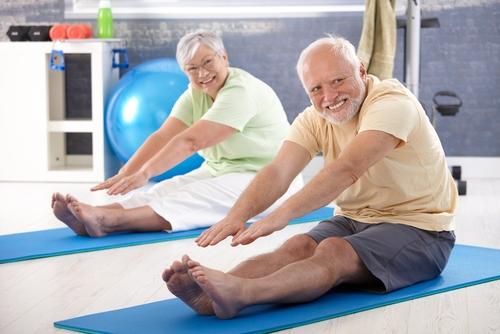
Do you think this doesn't concern you? Have you ever thought that your headaches, hypertension and even stroke could be due to cervical osteochondrosis? Rehabilitation doctor Alexander Shishonin assures that it is better to “twirl” your neck all your life than to be afraid of the occurrence of another disease.
Consequences of cervical osteochondrosis
The doctor claims that it is because of this disease that hypertension appears! Vertebrae cervical region undergo shifts over the years. As a result, compression of the neck vessels occurs. They, in turn, provide “nutrition” to the brain. As a result, there is a lack of oxygen, and the brain instructs the heart to pump blood much faster.
And, in the event that the heart is not completely healthy, the brain itself takes control of the entire process. It causes the blood vessels to contract strongly, increasing the pressure.
As a result, a change occurs in the composition of the blood, and sugar increases. Because the brain needs nutrition. And, if all supplies run out, a stroke may occur! Do you still doubt that you need to treat your cervical osteochondrosis? After all, trouble can come from where you don’t expect it at all! You may simply not even think about the fact that you are susceptible to such a serious illness, and just complain about neck pain all your life!
Shishonin's exercises
All these troubles are very easy to avoid if you do special exercises, which will remove the clamping of blood vessels by the vertebrae of the neck.
Clamping of blood vessels most often occurs due to nervous shock. When this happens, a spasm of the muscles of the neck and chest begins. Blood flow is disrupted and disc dysfunction occurs. The vertebrae begin to put pressure on the vessels.
To avoid unpleasant consequences and help patients with cervical osteochondrosis, Shishonin came up with a whole complex effective exercises, helping to get rid of neck diseases.
It is recommended to perform these exercises with additional training and diet. They are easy to perform even at home. After a month of implementation, you will be able to feel significant changes in your health. If this does not happen, it is recommended to consult a doctor.
Judging by the fact that you are now reading these lines, victory in the fight against inflammation of cartilage tissue is not yet on your side...
Have you already thought about inpatient treatment? This is understandable, because joint pain is a very dangerous symptom, which, if not treated in a timely manner, can result in limited mobility. Suspicious crunching, stiffness after a night's rest, the skin around the problem area is stretched, swelling in the sore spot... All these symptoms are familiar to you firsthand.
Arthritis of the ankle joint is a disease caused by an imbalance in the proportion of load on the joint and the stability of its components. When the first signs of this disease appear, you must immediately consult a doctor to establish the correct diagnosis and prescription. effective treatment.
The inflammatory process develops quite rapidly, motor activity is gradually limited, lifestyle is disrupted, temporary disability occurs, and in some cases even disability.
The main causes of ankle arthritis are:
- Heavy load of body weight and constant movement;
- Injuries (post-traumatic arthritis);
- An infection that enters the joint cavity along with the blood flow, as well as in the case of an open injury (in such cases we are talking about reactive arthritis);
- “Loss” of small crystals of uric acid in the area of the synovial membrane, due to gout or metabolic disorders;
- Autoimmune connective tissue diseases (gout, rheumatoid arthritis, systemic lupus erythematosus, psoriasis).
Classification
Arthritis has similar symptoms for all types and stages of the disease, so it is almost impossible to determine the type of disease without the help of a specialist. Arthritis of the ankle can be:
- Acute – in the presence of gout and infection;
- Chronic – in the presence of an autoimmune disease and osteoarthritis.
Most often, patients complain of severe pain in the affected joint, and there are cases of unilateral damage (in the presence of infection) and bilateral (in collagenosis), as well as migrating (in pseudogout and gouty arthritis).
Long-term arthritis can lead to joint fusion (ankylosis) and restriction motor abilities. This disease progresses progressively, with the onset of periodic periods of deterioration and improvement, but in any case, destruction of the joint occurs over time.
Manual therapy, ankle massage
After confirming the diagnosis, the patient must contact the attending physician or exercise therapy trainer to create an individual set of exercises. The specialist draws up a training plan depending on the patient’s condition, his physiological characteristics and the degree of advanced arthrosis.
Exercises should not bring discomfort or pain; all actions must be performed slowly and without sudden movements so as not to provoke a worsening of the condition.
During this period, gymnastics for ankle arthrosis improves microcirculation of lymph and blood, relieves muscle spasms, relaxes, and improves the patient’s well-being. It is advisable to carry out the exercises in a lying position.
Daily exercise helps improve the condition of the ankle joint and slows down degenerative processes in connective tissues.
Before training, be sure to warm up your joints and muscles with a small ankle massage. The exercise itself should consist of three blocks: light introductory exercises for warming up, the main block of exercise with increased load, and at the end, perform movements with reduced intensity.
An acute form of arthrosis of the ankle joint interferes with leading a normal lifestyle and brings pain and discomfort. Therefore, it is not recommended to do gymnastics at this time. A set of exercise therapy exercises for arthrosis of the ankle joint for recovery after an exacerbation:
- Sit on a chair and stretch your legs. Contract your feet, pull your toes towards you for 15 seconds, then relax for 3-5 seconds and repeat the movement 10 more times;
- Sitting on a chair, press your heels to the floor and rotate your feet in and out for 10 repetitions;
- Lunge with each leg alternately, shifting your body weight to the front leg and maintaining balance for a few seconds. Do 10-12 repetitions on both sides;
- Sit on a chair and roll from heel to toe and back. Perform the movement 10-30 times, focusing on sensations.
- Sitting on a chair, relax your legs and swing them back and forth in the form of a pendulum.
Strengthening the muscles and ligaments of the foot is necessary if the diagnosis of arthrosis of the ankle joint is confirmed. The following set of exercises is suitable for this:
- Do this while sitting or lying down circular movements feet first in one direction and then in the opposite direction;
- In a lying position, perform the “bicycle” exercise, alternately extending and contracting your foot;
- Place your feet together and perform shallow squats so that your foot is completely flat on the floor and does not come off. Repeat 10-12 times;
- Place a piece of cloth under your leg and sit on a chair. Try to crumple the flap without using your hands, using your toes;
- For the exercise you will need a plastic bottle, a roller or a wooden stick. Roll the projectile back and forth, kneading the entire sole of the foot.

Exercise therapy to restore damaged ankle ligaments must be done starting from the end of the first post-traumatic period (2–4 days). Any damage leads to hemorrhages and tissue swelling. Against the background of swelling or the development of a hematoma, compression of tissues and blood vessels increases.
Compression of the vessels leads to a violation of venous outflow. Stagnation occurs in the venous capillaries, gas exchange decreases. This condition is called hypoxia and implies a lack of oxygen. If there is very little oxygen, regeneration will not occur.
The use of exercise therapy for recovery after rupture of ankle ligaments leads to the fact that (the work of the lower leg muscles), due to which venous blood moves more actively to the heart. Another positive side is that during movement there is a large amount of nutrients in the joint, which are necessary for the regeneration of torn ligaments.
The effect of exercise therapy for ruptured or torn ankle ligaments occurs 2–3 weeks after the start of classes. The condition of the joint itself improves noticeably. Swelling subsides, the skin acquires normal tension, and its sensitivity improves.
When stretching, exercises begin earlier; dynamic exercises can be used. Do more approaches and increase the training time itself. When stretched, inflammatory manifestations are much less pronounced, which allows you to do exercises with a greater range of motion without fear of damaging the joint itself.
If the ankle ligaments are torn, exercises begin after the end of the acute period (up to 15–20 days). The time that has passed since the operation is also taken into account. In exercise therapy, work on distant structures (fingers, knees) is first prescribed.
Later, when postoperative phenomena subside (the wound heals, swelling subsides), small movements in the ankle joint are prescribed. The exercises are low-amplitude, mostly static. For ruptures, wearing an orthosis or other ankle braces is often prescribed. Read more about ankle bracing options here.

Sprained ankle ligaments
Patients with diseases of the lower leg or ankle joint quite often turn to medical institutions.
Ankle diseases:
- Therapeutic – arthrosis, arthritis of the ankle joint.
- Traumatic – rupture, sprain, ankle dislocation.
- Surgical - separation of ligaments with bone fragments, comminuted fracture, displaced fracture, habitual dislocation of the foot.
- Infectious – bacterial arthritis.
- Dermatological – psoriatic arthritis.
For all of the above diseases, therapeutic physical exercise for feet that restore not only the function of the ligaments, but also develop their elasticity.
Depending on the method of treatment of any disease of the lower limb, exercise therapy options for the ankle joint differ:
- IN supine position.
- IN sitting position.
- Standing.
- In move.
For comminuted fractures with displaced bone fragments, a three-level restoration system is used:
- Period of skeletal traction.
- Period of applying a plaster cast.
- The period after removal of plaster immobilization.
- Relieves pain and swelling in joints due to arthritis and arthrosis
- Restores joints and tissues, effective for osteochondrosis
This exercise is known to everyone: it is with it that the warm-up begins in physical education classes in kindergarten and school. It’s very simple: while doing household chores, move around the apartment, alternately stepping on different parts of your feet.
For example, walk to the kitchen on tiptoes and walk back to the room on your heels. Then you need to use the inside and outside of the feet. You need to perform the exercise barefoot. If the floor in the apartment is not very warm, you should wear thick socks or knitted slippers.
Sit on a sofa or chair with your legs extended and slightly elevated. Rotate your feet, keeping them suspended, first towards each other (inward), and then in the opposite direction. In order for the muscles and ligaments to receive the necessary load, you need to make 15-20 movements in each direction.
Walking barefoot is useful not only for strengthening the muscles and ligaments of the ankle. There are many acupuncture points on the feet, the impact of which strengthens the body’s defenses and improves the condition internal organs, reduces swelling and cramps, relieves the feeling of fatigue.
To really benefit from this activity, you need to choose the right surface for barefoot walking. A floor covered with linoleum or tiles made from pressed raw materials based on synthetic resins is unlikely to be suitable for this. It has been established that walking on the following types of surfaces has a positive effect on the body:
- grass or straw mats;
- bamboo mats;
- cork slabs;
- boards made from solid trees of various species;
- sand;
- small pebbles;
- dense short grass.
It may seem that cottage owners have more opportunities to use the right massage surfaces, but this is not the case. In any city apartment you can organize a training corner equipped with a mat or a shallow tray filled with smooth, medium-sized pebbles.
Ready-made massage mats that have several areas with different surfaces are also very convenient. When purchasing such a device, you should give preference to a rug made from natural, environmentally friendly materials.
Not all doctors treat manual therapy equally well. But in the early stages, with subluxations of the joint, this method will significantly alleviate the patient’s condition. The manual doctor will determine which method to use to treat the joint.
If arthrosis is at the initial stage of the disease and is accompanied by displacement of the head of the joint, then manipulation is performed to sharply release the joint and put it in its place. This is a quick and painful procedure, but improvement is seen immediately.
In more severe cases, a mobilization procedure is performed. It is done in a course over a year of treatment. The process itself is a slight traction of the joint, which helps relieve muscle tone and restore motor function of the ankle.
First stage
Complex conservative treatment
Comprehensive ankle rehabilitation after rupture and sprain is the most effective method quickly return to functionality. The peculiarity of complex activities is that they use not only physical education, but also physical procedures, proper nutrition, comfortable shoes, medications (if necessary).
The following set of rehabilitation measures is considered the most optimal:

Exist special complexes exercises that are aimed at restoring the ankle exclusively through physical education. These include:
- a set of exercises for the ankle according to Bubnovsky;
- a set of exercises for the ankle according to Evdokimenko.

Electrophoresis
Restoring an ankle after a ruptured or sprained ligament at home is no different from rehabilitation in special institutions. All exercises can be performed even without special simulators. The same applies to physiotherapy.
At home, it is best to use UHF for recovery. It is used throughout the entire recovery period. Physiotherapeutic devices from the following companies are used in medical institutions:
- Undaterm;
- Radmir;
- Arrow;
- Flow;
- Elfor.

The average price of a UHF device is 250 to 800 dollars. The price of electrophoresis machines starts at $100.
After confirming the diagnosis (X-ray of the axis of the leg and ankle, Salzman's X-ray, X-ray of the joint under load, CT-SPECT, MRI) and establishing the causes of the disease, the doctor may suggest a conservative treatment regimen, which will include:
- analgesic and restraining drug therapy - fast and slow acting symptomatic NPS drugs;
- therapeutic massage and physical exercise;
- physiotherapeutic procedures;
- dietary food;
- wearing orthopedic insoles and shoes, using special orthoses and support bandages.
Endoprosthetics of the ankle joint for arthrosis is one of the types surgical treatment. It only applies if conservative treatment didn't bring desired results, and other types of surgical operations are obviously unsuccessful - to normalize the sequence of movements and restore natural gait.

For ankle arthrosis, the following medications are used:
- injections and tablets– nimesulide, celecoxib, indomethacin, voltaren, movalis, diclofenac, tizanidine, nicotinic acid, trental, agapurine;
- ointments – traumeel-s, deep-relief, fastum-gel, diklak, butadione, bystrum-gel, finalgon;
- chondroprotectors– tablets and ointments based on collagen and glucosamine hydrolyzate;
- injection of synovial fluid polymer prostheses– synoviscus, adanthus, hyalurome, synocrom, hyalgan fidia.
Physiotherapy

Physiotherapy is traditionally included in the “domestic” complex of treatment of inflammatory pathologies, while the choice of a specific technique does not depend on the cause and type of joint damage, but is determined by its availability.
Relatively new techniques are cryotherapy (cold treatment) and homeosynia (injection of homeopathic medicine into acupuncture points).
Medical nutrition

Despite the fact that the effectiveness of treating degenerative-dystrophic changes in cartilage tissue by following special dietary recommendations has not been 100% proven, nevertheless, a diet for arthrosis of the ankle joint is included in the complex therapy of this disease.
What to do?
First, it is necessary to normalize the volume of clean water consumed per day (min in l):
- women = body weight x 0.03 0.3;
- for men = body weight x 0.04 0.4 l.
Secondly, you should know which foods and dishes are “needed” for inflamed joints, and which ones should be avoided. The instructions are given in the form of a short table compiled from the recommendations of the nutritional textbook by A.Yu.
Baranovsky, and individual nutrition for ankle arthrosis with a daily menu and the required amount of kcal, taking into account the entire “bouquet” of existing diseases and daily physical activity, can only be compiled by a certified nutritionist.
| Shown | Not recommended |
| Fish fat | Dried cereals (except rice and wheat) |
| Evening primrose oil | Legumes |
| Pasture raised beef | Frozen fruits and vegetables |
| Mutton | Conservation |
| Premium hard cheeses | Smoked meats |
| Turkey | Spinach, sorrel, cauliflower, mushrooms |
| Low-fat natural yogurt | Marinades and seasonings |
| Unrefined vegetable oils | Pickles, salt |
Massage

Massage is not a very complicated procedure, but multi-stage. You can entrust massaging a sore joint to a specialist without a higher medical education, but only to a graduate of massage therapist courses.
The massage procedure should last at least 30 minutes and include locations in the following order:
- all types of massage techniques on the calf muscle in the following positions: lying on the back and on the stomach;
- massage of the anterior surface of the ankle;
- elaboration back surface;
- all types of passive movements, followed by massage of the calf, Achilles tendon, outer part of the lower leg;
- rubbing the foot.
Exercise therapy for arthrosis

The selection of physical exercises, their varieties, and especially the number of repetitions of each movement and the dosage of daily loads on the joint is the prerogative of the attending physician. In addition to the stage of development of arthrosis, it is necessary to take into account the reasons for its occurrence and the type of deformation to which this pathology led.
We present to your attention basic complex“general” foot exercises used in the German orthopedic clinic Gelenk-Klinik for the rehabilitation of athletes suffering from secondary arthrosis due to chronic external instability of the ankle joints.
READ ALSO: Essentuki Water - New, mineral water, mineral springs, indications for use, Essentuki mineral water, recommendations, treatments
| Image | Description |
|
| The exercise is performed in a sitting position and consists of 2 parts:
Repeat the cycle at least 3 times. |
|
| This simple exercise will help to effectively stretch the back group of muscles and tendons of the calf, strengthen the front tibialis muscle, superior and inferior retinaculum of the extensor tendons. Place your affected leg on the “cushion” and wrap one end around your foot over your toes (a). Move your fingers away from you, overcoming the resistance of the tape, which is dosed with your hand (b). Repeat 10 times. |
|
| Strengthening exercise deep muscles shins, and restoring the elasticity of their tendons, is performed in the following way. It is necessary to take the starting position with a stretched elastic band (1). Overcoming resistance, rotate your foot to position (2), and then slowly return your leg to the starting position. Do the exercise in 3 sets, 4-6 movements each. |
|
| To strengthen the peroneal muscle and the group of flexor and extensor muscles of the foot, a loop hold is performed elastic band, which can be attached to the cabinet leg. Before you start performing the movement towards yourself, the foot should already be in a tense state. The leg is in the starting position for 3 seconds, and then the same amount of time on itself. Number of repetitions until fatigue. |
|
| Fasten the elastic band loop as shown in the photo. While tensioning the tape, passively turn your foot outward (2), then, overcoming the resistance of the stretched material, return it to its original position. The exercise returns the fallen arches of the foot to their natural position by strengthening the deep peroneal muscles. Repeat 10-20 times. |


The basics of physical therapy for the ankle
Exercise therapy is of great importance in the treatment of ankle injuries. Its positive effect on the osteoarticular apparatus is undeniable, because only through movements can the normal function of all joint structures be restored.
The basis for the treatment of many injuries is early activation of motor function. The time to begin exercise is determined by the type of injury sustained and previous treatments.
In cases of surgical treatment of fractures or immobilization, development of the ankle joint begins somewhat later. However, you need to perform exercises for other joints and muscles, which will indirectly improve blood circulation in the affected area.
Performing exercise therapy during the period of immobilization for ankle fractures ensures normal blood circulation in the affected areas, maintains muscle tone, and preserves the function of healthy parts of the limb.
- In all unaffected joints: knees and hips on both sides, as well as in the ankle healthy legs.
- In the toes of the diseased limb - flexion, extension, extension.
- Raise and lower the injured leg.
- Rotational movements of the limbs in and out.
- Abduct and adduct the straight limb.
- Raise the affected limb with outward and inward rotation.
- Tension of the muscles of the sore leg in isometric mode.
Walking with support on an immobilized limb is of great importance. This is carried out only after the permission of the attending physician, gradually increasing the duration - from several minutes to an hour per day.
You should not attempt to stand on your affected leg on your own without medical advice - this may disrupt the healing of the fracture.

The exercises that can be performed after removing the plaster cast are significantly expanded in scope. They are designed to restore the function of the limb, as well as prevent the development of flat feet, when it is necessary to strengthen the muscles of the foot. In addition to those described earlier, the following exercise therapy exercises are performed in a sitting position:
- Flexion and extension of the foot.
- Rocking movements of the foot to relax the muscles.
- Place your feet on your toes, pushing your heels out and vice versa.
- Walk while sitting, rolling your foot from heel to toe.
- Rotational movements of the foot.
- With your foot on the stick, roll it in the center, with the outer and outer edges of your foot.
- Stretch forward with the toe of your straight leg.
- Leaning on the toe, make springing movements with the heel.
- Grab a small object with your toes and hold for a few seconds.
During this period, it is necessary to bandage the ankle with an elastic bandage without getting out of bed, and when performing gymnastics it is removed.
Classes are carried out carefully so as not to provoke pain. Gymnastics are performed 2-3 times daily.
In order to reliably strengthen the muscles and ligaments of the affected ankle, as well as completely restore its function, it is necessary to continue exercise therapy for a month after the injury. During this period, the healing of the fracture ends, so you can apply more force to the ankle and increase the duration of the gymnastics. The following exercises are recommended, performed with support on a gymnastic wall or the back of a chair:
- Rolling from toes to heels and back.
- Transfer body weight from one limb to another.
- Squat on your entire foot and toes.
- Walk on toes, heels, outside feet, side steps.
- Lunges forward with the affected limb.
- Standing on the stairs on your toes, perform springing movements, lowering your heels as much as possible.
All elements of therapeutic exercises for ankle joint injuries must be performed in strict accordance with medical recommendations - only a specialist will indicate the required range of movements, their frequency and duration.

First, the classes are conducted under the supervision of an instructor, and then, after training, the patient can perform them at home. A properly selected set of physical therapy will become the basis for recovery from ankle injuries.
How to strengthen ankle joint
At various stages of treatment, special exercise therapy complexes for the ankle are used. Classes are carried out gradually; sudden movements should not be allowed.
In order for ankle development after a fracture to be successful, a special set of exercises is selected at each stage of treatment and recovery. Each of them has its own specifics, but they have one thing in common: sudden movements of the injured leg cannot be made.
At the first stage of recovery, when the fractured leg requires complete rest, the patient engages in physical activity without the participation of the injured leg.
Such classes are needed at the first stage of recovery for the following purposes:
- Improve blood flow throughout the body to ensure adequate nutrition of organs and tissues;
- Prevent the development of stagnant processes;
- Prepare the ligamentous and muscular apparatus for subsequent classes.
At the second stage, when the plaster is removed, physical activity necessary for recovery muscle strength and reducing atrophy.
At the third stage, a period of active training begins. The ligaments and muscles prepared in the previous stages are involved in intense physical activity. At this stage, exercises on an exercise bike, treadmill, and in the pool are added to the usual exercises.

Let's look at the features of exercises for developing the ankle joint at each stage.
The first stage of rehabilitation is a difficult and responsible time: the patient must combine careful treatment of the broken leg and minimal stress.
The first exercises, which are started 3-4 days after applying the plaster, are performed lying down. When performed, they activate the muscle tissue located near the fracture.
- Consistently tense the muscles of the buttocks, thighs and calves up to 20 times;
- Move your toes, squeezing and unclenching them for 1-2 minutes.
After 7-14 days (the specific period for when to start more active exercises is prescribed only by the doctor), they move on to more active training. Their task is to step on the leg with a broken tibia, using it as a support when walking.
- Sit on a chair. Try to straighten the knee of the affected leg so that the limb takes a position parallel to the floor. “Return” your foot to the floor. Do 5-6 approaches.
- Without changing your position, pull the knee of your broken leg towards you with your hands. Lower your leg. Do 5-6 approaches.
- Stand with your hand on the back of a chair. Move your injured leg to the side. Place it on the floor. Repeat the movement 10-15 times.
- Move your injured leg back. Put it down. Repeat the movement 10-15 times.
- Smoothly “point” your leg forward. Put it down. Repeat the movement 10-15 times.
- Repeat the previous exercise. But after pointing your leg forward, fix it in this position for at least 3-5 seconds. Place your foot on the floor. Repeat the movement 10-15 times.
During training, listen carefully to how you feel. Fatigue and discomfort in muscle or joint tissue are evidence that exercise should be stopped. If the pain is minor, you can reduce the number of approaches or range of motion. At the beginning of training, it is better to take short breaks after each exercise performed.
As soon as the patient's plaster is removed, it is necessary to begin working on the injured joint itself. At first they practice every other day. After the muscles get used to the load, they move on to daily training.
The main position at this stage is sitting on a chair. It is important to remember that 2 legs are involved in the exercise: the sick one and the healthy one. All movements are repeated for 2-3 minutes.
- Without lifting your feet from the floor, alternately “roll” from heel to toe. The movements are made first with two legs, and then with each in turn.
- “Rise” on your toes and stay in this position for 2-3 seconds. Lower your entire foot.
- Press your heels together. Lift your socks up while turning them to the sides.
- Press your socks together. Lift your heels up while simultaneously turning them to the sides.
- Focus on your toes, slowly “rotating” your heels from right to left. Then change the direction of rotation.
- Focus on your heels, slowly “rotating” your toes from right to left. Then change the direction of rotation.
- Extend your legs forward. Make rotational movements with your entire foot from right to left. Then change the direction of rotation.
The goal of the third recovery stage is to restore the functionality of the broken tibia. Therefore, all exercises involve active walking. To “protect” the still weak joint, all loads are performed using an elastic bandage that secures the ankle joint.
- Walking on your heels.
- Walking on toes.
- Heel-to-toe walking.
- Standing on the canvas, use your toes to “gather” it under your feet.
- Roll with your feet plastic bottle.
At this time, it is important to take daily walks, go up and down stairs, attend massage sessions, physiotherapy, and maintain a proper and balanced diet.
Complete restoration of the functioning of the ankle joint after a fracture is impossible without proper rehabilitation.
Forced immobility for several weeks leads to partial atrophy muscles and weakening of the ligamentous apparatus.
Immobilization after a fracture - rigid fixation of bones with a plaster splint after their preliminary alignment ensures retention of the joint in correct position.
In case of complex injuries with the formation of fractures, surgical treatment is carried out - strengthening the bone fragments with metal screws or plates.
A plaster splint is applied for a period of 1 to 3 months, depending on the type and complexity of the fracture.
Measures to strengthen the joint tissues must be taken when the leg is in a cast - already on the second or third day of immobilization. The first exercises after an ankle fracture do not involve active movements and the participation of the damaged ankle. The muscles only adjacent to the affected area are involved. You can perform them while lying in bed.
- Tighten and relax the gluteal, thigh and calf muscles. Repeat alternately with the sore and healthy legs 10-20 times.
- Squeeze and unclench the toes of both feet for 1 minute.
Exercises after removing the cast
The article describes the need for physical therapy in the complex treatment of fractures. Exercises used for fractures of the ankle joint (articulatio talocruralis) are described.
Treatment of fractures consists of several stages. First of all, restoration of bone integrity and healing of damage is carried out.
After this, rehabilitation treatment is prescribed, aimed at preventing complications and maintaining motor function. Exercise therapy after an ankle fracture is one of mandatory stages rehabilitation treatment.
Any fracture is a violation of the integrity of the bone and adjacent structures. This leads to disruption of the functioning of the part of the body where the fracture occurred. With fractures of the articulatio talocruralis, the function of the lower limb is affected. The person moves with difficulty or cannot walk at all.
After a fracture, a person has difficulty walking
Even after healing of the fracture, residual effects remain associated with damage to soft tissues and prolonged exposure to a forced position. After removing the plaster cast, motor function suffers significantly and movements cause pain.
Physiotherapy This period eliminates attempts to step on the sore limb or rotate the damaged joint. Movements should be smooth and careful.
Exercises in a sitting position:
- Keeping your back straight, place both feet on the floor. Bend the knee of the affected leg, stretching it parallel to the floor. Return to starting position.
- Raise your leg without straightening it, pulling your knee as high as possible, then put it back on the floor.
Standing on the floor:
- For support, you can use the back of a chair, table, crutch or wall. Slowly and carefully move the injured leg to the side and place it back on the floor.
- Perform the same exercise, holding your leg at the extreme point of the lift for a few seconds.
- Take your leg back, trying to lift it as high as possible, and return to the starting position.
- Perform the previous exercise with the leg held in the extreme position.
- Swing your leg forward smoothly, then return it to the floor.
- Perform a circular swing with your leg: take it back, describe a semicircle in the air, moving it forward.
Place your foot on the floor. Repeat the movement in the opposite direction.
Perform each exercise 10-15 times.
The above complex helps strengthen muscular frame legs, helps improve blood flow and tissue nutrition, reduce swelling.
Therapeutic exercises for a broken ankle require careful attention to your well-being. If there is severe pain in the area of injury, the exercise should be postponed or its amplitude reduced.
When practicing for the first time, you don’t need to set strict limits for yourself and try to complete the entire complex at all costs, trying to achieve the maximum number of repetitions through force. Appearing fatigue or physical pain is a sign that you need to rest.
It would be optimal to take a short break after each exercise, otherwise not only the sore leg will get tired, but also the healthy leg. After some time, provided you confidently maintain your balance, you can begin to perform exercises without additional support.

Physical therapy after an ankle fracture
After removing the cast, the leg does not look in the best possible way - skin pale or bluish, swelling in the ankle area, thinner calf muscles. But the main problem of a limb freed from shackles is stiffness.
Therapeutic exercises after an ankle fracture are necessary to strengthen the ligaments, restore normal blood supply, tissue innervation, and return a healthy gait without lameness. After removing the cast, attention is directed directly to the ankle joint itself.
Classes are conducted under the guidance of a trainer in a special group.
The intensity of the load is always calculated individually, based on the severity of the injury, severity of pain, physical condition, and body weight of the patient.

Exercises after an ankle fracture in the first days should be performed while sitting on a chair, this will provide a more gentle load on the fracture area. Shoes must be removed from your feet during exercise. Both legs should work synchronously. Each movement must be repeated for 2-3 minutes.
- Place your feet on the floor parallel to each other. Raise your toes up and down without lifting your heels off the floor. Perform rhythmically, first with both feet simultaneously, then alternately.
- Raise your heels as high as possible and lower your heels, resting on your toes. Alternate simultaneous and alternate execution.
- Smoothly roll your feet from heels to toes and back.
- Feet stand side by side on the floor, heels pressed together. Raise your toes up while simultaneously turning them to the sides and return to their original position.
- Thumbs parallel feet standing on the floor pressed against each other. Raise your heels up, spreading them apart.
- With your toes resting on the floor, make circular movements with your heels clockwise and then counterclockwise.
- Perform similar movements with the toes of your feet, resting your heels on the floor.
- Straighten your knees, stretching them forward. Rotate your feet in different directions, pull your toes towards you, clench and unclench your toes.
Having mastered the complex in a sitting position, you can move on to performing it standing, holding onto a support. To those listed physical therapy exercises after a fracture, as the ligaments and tendons strengthen,
READ ALSO: Unicondylar knee replacement
New ones are gradually being added:
- Walking on your heels, then on your toes, on the outside, on the inside of your feet.
- Steps with rolls from heel to toe.
- Spread a thin towel or napkin on the floor. Stand on its edge with bare feet.
Using your fingers, grab and gather all the fabric under your feet.
- Use a plastic bottle, rolling pin or other cylindrical object with a smooth surface. Roll the training apparatus with your feet on the floor back and forth.
- Place a small rubber ball on the floor, place your foot on top and turn it from side to side, pressing with your toe, heel, and the outside and inside of your foot.
In order to relieve fatigue, prevent or eliminate signs of swelling, before classes and during rest after them, apply to the sore leg for 30-40 minutes. It is useful to lay horizontally on the armrest of a sofa, chair or armchair.
Along with therapeutic exercises, massage, physiotherapy, and baths are used to restore and strengthen the health of the joint. It is necessary to follow a diet - eat foods rich in calcium. In the absence of contraindications, you can introduce exercise on a treadmill and jumping.
To fix and support the ankle, you must use a medical orthosis or an elastic bandage.
The total duration of therapeutic rehabilitation sessions ranges from 1 month to six months.
The success of rehabilitation depends on the severity of the injury, the age of the patient, and the presence of concomitant bone diseases.
Except special classes in the group you can find a lot of ways to develop a leg after an ankle fracture.
Regular walking during street walks, going up and down stairs are excellent physical exercises that help cope with the residual effects of injury.
Cycling, swimming, and working with a foot-powered sewing machine have an additional training effect.
Ankle fracture requires correct selection shoes Women will have to give up stilettos and high heels or platforms. It is necessary to use orthopedic insoles for several months.
Ankle stiffness and soft tissue swelling in the injured area, lameness and low exercise tolerance are common Negative consequences injuries. To prevent complications and restore normal motor activity of the foot, rehabilitation after an ankle fracture is important, which is prescribed and developed by a traumatologist or rehabilitation specialist.
Ankle fracture is considered one of the most common injuries lower limbs. The ankle is the tubercle in the lower third of the leg bones. The medial malleolus is located with inside ankle joint and is a bony protrusion tibia.
The lateral malleolus is located on the outside of the ankle and, accordingly, is the bony protrusion of the fibula. The junction between the bones of the lower leg and foot has a weak ligamentous apparatus, and at the same time bears a large load.
The risk of fractures in the ankle area is especially high when playing sports, wearing uncomfortable high-heeled shoes, or being overweight.
Schematic location of an ankle fracture
An ankle fracture can be open or closed. Diagnosis of a fracture and determination of its type is carried out using radiography. An open fracture is the most severe type of injury in which the bones are damaged soft fabrics and exit through the wound surface.
A closed feather does not form an open wound, it can be with or without bone displacement. A fracture without displacement is less likely to cause complications and heals within 2-3 weeks. An injury with bone displacement takes longer to form a callus.
Sometimes surgical intervention is performed to give the anatomically correct localization of bone fragments. These circumstances extend the recovery period.
After applying a cast to the limb, the patient is recommended to rest and limit physical activity for 1-2 weeks after the injury.
Rehabilitation measures are carried out after the end of the period of immobilization of the injured ankle joint.
To restore lost functions and develop the ankle, which has been in a stationary position for a long time, massage, physiotherapy and therapeutic exercises are prescribed.
The doctor may prescribe some physical therapy exercises before removing the plaster cast if the fracture healing dynamics are positive.

Preventive exercises after plaster removal
Breathing exercises for lower leg injury
The choice of exercises depends on the injury received and the time that has passed since it. With sprains, people with average physical training can perform two sets of exercises. The first is used for warming up, the second is used to improve blood flow.
The first group includes the following exercises:

The second cycle includes exercises with support on the foot. The following exercises are used:
- Lying on your back, your legs should be raised at an angle of 45–50 degrees and placed with even feet on the wall. Alternately change the position of your foot from toe to heel, lightly resting on the wall to increase the load. Select the duration of the load individually, based on your physical characteristics, on average 45–60 seconds per approach.
- Lying down, legs straight, heels resting on the floor. Start turning your foot clockwise and then counterclockwise, using your heel as a pivot. It is necessary to perform 15–20 times, 2–3 approaches.
- Standing at a support, you need to rise with both feet on your toes to the maximum height. It is necessary to do 5–10 times, over time the amount should increase to 10–15 times.
In addition to the basic exercises, you can strengthen the training of the ankle ligaments with exercises with props. For quick recovery use the following complex:
- Exercise with a gymnastic band. Sitting on the floor with with outstretched legs, put a wide rubber band on the ankles. Slowly begin to abduct the legs to the sides, stretching the elastic band. You need to return your legs to their original position just as slowly. Perform 10–12 times, 1–2 approaches.
- Exercise with a rolling pin or rubber ball. Place a rolling pin under your feet and begin to swing the dough on the floor impromptu using foot movements. Using your foot, you need to roll the ball from your toes to your heel.
In people with good physical fitness, recovery is a little faster. 20–30 days after an ankle ligament injury, they begin active walking on their toes and heels, changing the supporting part every 1–2 minutes.
There is evidence that it is possible to walk on the inside or outside of the foot. It is possible to walk on the outside of the foot when there is a medial ligament injury. On the inside of the foot - when there is an injury to the lateral ligament.
This is another exercise, familiar from childhood, that strengthens the ankle. To complete this, you will need small hard objects (pebbles, glass balls, buttons, etc.) and a low container. Sitting on a chair, you need to collect objects from the floor, grabbing them one at a time with your toes, and transfer them to a basin or box.
The exercise can be performed standing on one leg (legs should be changed periodically). In this case, it will serve not only to improve the condition of the ankle, but also to train the vestibular apparatus and coordination of movements.
The ankle is a complex bone structure, since it is built not from two, but from three bones: the tibia (fibula and tibia) and the talus. The lower surfaces of the tibia form a kind of fork, which covers the block of the talus bone.
This connection acts as an axis around which the foot moves. The lateral ends of the “fork”, that is, the processes of the tibia bones, are called ankles. The articular surfaces are covered with hyaline cartilage tissue, which acts as a shock absorber.
To ensure that the bones do not fall apart, stay together and function, a whole system of ligaments is involved.
They can be divided into three groups:
- Fibers that bind the shin bones together and prevent them from turning.
- The outer lateral fibers that hold the joint together from the outside.
- The deltoid ligament firmly connects the talus, calcaneus, and navicular bones of the foot to the medial malleolus.
“Ribbons” of ligaments help the foot move and at the same time limit its movement. Muscle tissue allows the feet to perform a variety of movements.
They are also divided into three groups:
- The flexors are located on the back of the ankle.
- The extensors are located on the front of the joint.
- The arch supports and pronators are responsible for moving the foot in and out.
The mobility of the joint also largely depends on the Achilles tendon, the largest in our body. It stretches from calf muscle to the heel bone. The tendon provides additional protection to the ankle. At the same time, it belongs to the most vulnerable areas and is often injured.
Did you know? The mobility of the ankle changes with age: in young children it bends more easily towards the back of the foot, and in adults it bends more easily towards the sole of the foot.
The ankle joint is surrounded by a dense network of blood vessels. Thanks to many veins and arteries, an uninterrupted metabolic process takes place in it.
The ligament system is quite strong; it reliably holds the bones in the joint and allows them to move properly. But one sudden and incorrect movement of the foot, for example, tucking it, and the ligament attachments may not withstand the unexpected load. The joint twists unnaturally and the ligaments become damaged.
The most common causes of ankle injury:
- walking in high heels;
- excessive tension on the joint sports training;
- heavy physical labor;
- incorrect landing during a jump or fall;
- the foot turned up;
- moving on uneven or slippery surfaces;
- striking the leg.

As a result, various joint elements may be damaged: bones, muscles, ligaments and tendons.
Based on the type of damaged tissue, the following ankle injuries are distinguished:
- bruise - violation muscle tissue;
- dislocation - damage to bones, in which their position is completely or partially displaced;
- fracture - damage to bones as a result of a fall or blow;
- sprained or torn ligaments.
There are 3 degrees of severity of ankle injuries:
- A minor injury (for example, a sprain) in which there is a rupture of the joint fibers, but the stability and functionality of the joint is not lost.
- The ligaments are partially torn, which causes severe pain, but the joint functions.
- The ligaments are completely torn, which causes acute pain with any movement, swelling and hemorrhages are observed, the joint does not function. This serious injury, the treatment of which requires urgent treatment, and the recovery period is delayed.
Did you know? A person whose mass is 60 kg, taking one step, puts a load on his feet and ankles of about 240 kg. During the day, his legs can carry a load of up to 2.5 million kg.
Frequent leg injuries can be the result of a weak ankle joint.
You can recognize an ankle that needs strengthening by the following symptoms:
- cases of sprains after a jump;
- after a long walk, a feeling of pain in the third and fourth toes;
- frequent twisting of the foot in high-heeled shoes;
- external manifestations: in a standing position, it is visually noticeable that one or both feet are skewed inward.
Breathing exercises necessary to prevent blood stagnation in the lungs and prevent the occurrence of pneumonia. This condition can develop with prolonged bed rest. The most best exercise from the breathing exercises complex this is exhaling through a straw into a glass of water.
It is correct to perform various turns and bends, which will not only improve brain function by expanding the vessels of the spine, but also strengthen the muscles of the anterior abdominal wall.

Exercises with a tourniquet for an Achilles injury
Typically, after surgery for tendon repair, a blind cast is applied for 3 weeks. It is above the knee joint, so it is almost impossible to work out the sore leg during this period. In order to increase the overall muscle tone in the body, you need to do the following exercises:
- Pull-up.
- Push-ups.
- Arm movements with dumbbells.
- Rotations and bends of the body with dumbbells in hands.
- Slow but amplitude movements in hip joint sick and healthy legs.
- Breathing exercises (Quick inhalation and slow exhalation for 10-15 minutes).
A set of exercises after 3 weeks of Achilles tendon injury:
- Active flexion and extension in knee joint.
- Maximum range of motion in the hip joint of the affected leg.
- Lifting your legs up while lying on your side.
- Description of a circle with the affected leg in the air in various positions of the body: sitting, lying, standing.
A set of exercises after removing immobilization from the Achilles:
- In the water we work with all joints of the leg: bending, unbending, drawing circles.
- We step on our feet as much as possible in the water.
- At home, we roll a wide bottle with both feet at the same time.
- Exercise “heel-toe” - we take turns touching the floor with the heel and toe.
- Walking up the steps. First we walk on steps with a height of 5-10, then gradually increase them.
- Exercises for static resistance. We rest our hands on the leg and prevent it from making any movement in any direction.
Towards the end of the recovery period, you can begin to do deep lunges, squats and jumps. But it is immediately worth noting that only symmetrical work of both legs is the key to success in rehabilitation after injury.
Physiotherapy procedures are prescribed immediately after the end of the period of immobilization of the ankle joint. The impact of physical factors on the fracture area improves blood flow, activates metabolic processes, reduces swelling of soft tissues, and has an analgesic effect.
Contraindications
Although physical activity is essential for recovery from a broken ankle, it is not for everyone.
The following pathological conditions are contraindications to exercise therapy:
- Inflammatory and purulent processes in the body;
- Diseases in the acute stage;
- Pregnancy;
- Unstable compound fracture;
- Mental illnesses;
- Malignant formations;
- Bleeding from all locations.
Exercises with a rubber band
A pharmaceutical rubber bandage is one of the most successful devices for strengthening training. With its help, you can do exercises that have a positive effect on the muscles and ligaments of the ankle:
- sitting on a chair, place your feet close to each other and tie them with a bandage, securing the knot on top. Now place your heels on the floor and lift your toes. In this position, try to spread your socks to the sides several times, stretching the bandage;
- secure the bandage in the same way as in the previous exercise. Cross your feet, place your heels on the floor and again stretch the bandage several times, trying to move your toes as far apart as possible;
- secure the bandage knot to a support (such as a cabinet leg). Place the toe of your foot into the resulting loop (the heel should be on the floor) and pull it towards you several times, overcoming the resistance of the bandage. The movements can be performed with each leg separately or with both legs together.
Jumping rope is considered an excellent exercise for the ankle. Experts recommend jumping without bending your knees: in this case, the ankle joint receives the necessary load.
Weakness of the muscles and ligaments of the ankle is fraught with frequent injuries lower legs. If such a problem occurs, you need to choose the right shoes for leaving the house. For a regular walk or work, hard boots that keep their shape well are suitable.
Orthopedic insoles are also very useful in this case. For active rest You should wear high-top sneakers that hold the joint in a fixed position. Of course, in case of an ankle injury, you should consult a doctor and discuss with him not only treatment issues, but also the choice of strengthening exercises.
Strengthening and prevention

A weak ankle is the cause of most fractures and other ankle injuries. To avoid dangerous injury, an important joint should be strengthened. You can do this with the help of exercises:
- Jump rope regularly. It is better to do this without shoes: the foot will receive maximum load.
- Run on your toes.
- Collect small objects from the floor with your toes: beads, beads, etc.
An important component of the prevention and strengthening of muscle and bone tissue- nutrition. Include calcium-rich foods in your diet and control your weight: excess weight will negatively affect the condition of the lower leg. Drink at least 2 liters of water per day and drive active image life.
Often the ankle is injured during sports, as well as at home due to one’s own carelessness.
The type of damage depends on the degree of impact of the mechanical factor. This also determines the symptoms of the pathology, which in each case will have their own characteristics. But even a minor injury can cause significant inconvenience to the patient, because walking and support function are impaired, without which it is impossible to imagine everyday life.
Osteochondrosis is a chronic disease of various parts of the spinal column, in which degenerative and dystrophic changes occur in the intervertebral discs. This disease most often affects men aged 25-60 years. The disease is typical for both young people and old people. Women get sick less often, and their symptoms are not as obvious. At the initial stage of the disease, it is possible to achieve stable remission and get rid of pain and discomfort for long years.
What are the types of osteochondrosis by localization?
The classification of spinal osteochondrosis has many forms and criteria. They differ in the location of the affected area and severity. All types of osteochondrosis are pathologies that are fraught with destructive changes in cartilage and connective tissue.
The most common and understandable classification for the common man, far from medicine, is based on the location of the lesion:
- Cervical osteochondrosis - it is characterized by occipital, cervical and interscapular pain syndromes. This disease has been increasingly diagnosed in young people over the past decade.
- Osteochondrosis thoracic The spine is very rare, since it has more discs than in the cervical and lumbar combined, the discs are smaller and thinner. Part of the load is taken by the ribs. Therefore, degenerative and dystrophic changes rarely occur in the intervertebral discs of the spine.
- Lumbar osteochondrosis most often occurs in people over forty years of age from a sedentary and sedentary lifestyle. It is characterized by severe pain in the lower back, pinching of the nerves and arteries of the abdominal organs, and many concomitant diagnoses of internal organs.
Stages of the disease
Depending on the speed of the course, the degree of degenerative changes, and the manifestation of symptoms, four stages of the disease can be distinguished:
- The initial stage is characterized by instability of symptoms. The patient is not yet experiencing serious pain. Occasionally, shooting pain in the affected area of the back is possible.
- The second stage is characterized by a significant deterioration in the interaction of the vertebrae due to the progression of degenerative processes in the intervertebral discs. The development of the disease at this stage is provoked by incorrect physical activity, sedentary and sedentary lifestyle.
- At the third stage, intervertebral hernia may begin to develop (some patients avoid this fate). In some cases, severe back pain occurs. Concomitant diagnoses are developing that in one way or another affect all systems of the body. Symptoms vary depending on the location of the hernia and the location of the chondrosis.
- The fourth stage is the most difficult. The patient is unable to lead a full life and enjoy freedom of movement. Almost all postures occupied by the body bring acute pain at the site of osteochondrosis. At this stage there may be talk about assigning a disability to the patient.
Causes
Osteochondrosis brings a lot of suffering, worsens the quality of life, and can lead to complete or partial immobilization of the patient and disability. What are the causes of this disease? They are as follows:
- Sedentary lifestyle, static sitting or lying in one position. The muscles atrophy, the vertebrae wear out the cartilage and discs between each other. This process can last for years, but will inevitably lead to osteochondrosis.
- Modern boys and girls go to the gym and perform exercises with barbells and dumbbells, the improper execution of which leads to chronic diseases of the spine. For example, the incorrect technique for performing the exercise “squats with a barbell” presented surgeons and orthopedists with tens of thousands of young osteochondrosis and scoliosis patients in the acute stage.
- People who, due to their professional activities, are forced to sit at a table in one position for many hours in a row are also at risk. According to WHO statistics, office workers make up the lion's share of patients with osteochondrosis.
The main symptoms of cervical osteochondrosis
Gradually they lose fluid and become brittle. As a result of this process, compression of the veins and arteries occurs. The nutrition of the brain is disrupted and subsequently the death of its cells. Cervical osteochondrosis has the maximum negative effect on the area of the brain responsible for the activity of the heart muscle. The optic or auditory nerve is often compressed, leading to vision loss and hearing impairment.
Types of cervical osteochondrosis:
- radicular, or so-called “cervical radiculitis” - it is characterized primarily by severe pain in the back of the head, neck, and head;
- irritative-reflex - discomfort in the chest, back, numbness of the fingers;
- cardiac, which is characterized by symptoms of cardiovascular diseases.
Only an orthopedic doctor or surgeon can make an accurate diagnosis. Regardless of the type of cervical osteochondrosis, the treatment will be almost identical.

The main symptoms of osteochondrosis of the thoracic spine
The rarest type of disease. Symptoms of osteochondrosis of the thoracic spine are characteristic of many other diseases. An inexperienced doctor can often diagnose arrhythmia, angina, cardiomyopathy (before receiving ECG results), pancreatitis or even gastritis. Only an experienced orthopedist or spinal surgeon can provide accurate information about the type of osteochondrosis of the thoracic region.
With a pronounced pathological process, a disruption in the functioning of areas occurs spinal cord. The main signs of osteochondrosis of the thoracic spine:
- Dorsago - sharp, unbearable pain in the chest, a feeling of lack of air, the patient feels as if he is suffocating or his heart rate is abnormal.
- Dorsalgia - the patient experiences less discomfort than with dorsalgia. The pain is less obvious and radiates to the hands and fingers (which is why doctors often confuse the symptoms with diseases of the cardiovascular system).

Symptoms and lumbar spine
The most common type of osteochondrosis is lumbar region. This is due to the fact that it is the lower back that experiences the greatest load in the process of daily life.
In the first stage, the main symptom is a slight stretching in the lower back. People rarely attach importance to such minor ailments. As wear and tear increases, negative symptoms also increase. In particular, the patient can no longer lie or sit in one position for a long time due to pain.
At the third or fourth stage of lumbar osteochondrosis, the following occur:
- lumbago - sharp and sudden pain, the result of which can be partial immobilization of the patient;
- sciatica - unilateral infringement of the sciatic nerve;
- radiculitis - compression of nerve endings in the lumbar spine;
- weakness of the lower extremities, muscle atrophy, venous congestion, dysfunction of sexual function and the functioning of the abdominal and pelvic organs are also often observed;
- radiculoischemia - failure of blood circulation as a result of compression of blood vessels.

Generalized osteochondrosis
This type of disease affects not only the spine, but also the joints. Most often the shoulder and hip. Symptoms of generalized osteochondrosis are neurological in nature, in rare cases with damage to internal organs due to impaired blood circulation.
In the hip joint, the disease manifests itself as changes in cartilage tissue and drying out of the interarticular fluid. Over time, the joint partially and then completely loses its former mobility.

Polysegmental osteochondrosis
Manifestations of the disease are not in one, but in several parts of the body. For example, there is often a combination of damage to the third and twelfth vertebrae - in this case, osteochondrosis will be considered polysegmental.
Therapy in this case will be exactly the same as for standard signs of cervical or thoracic osteochondrosis. Magnetic therapy and other physical procedures have proven to be excellent for exacerbation of pain and discomfort due to polysegmental chondrosis. Now you know what types of spinal osteochondrosis exist.
Drug treatment of osteochondrosis
Pharmacological agents to alleviate the symptoms of osteochondrosis can be divided into the following groups:
- Anti-inflammatory medications - to relieve symptoms of pain and swelling in the affected area. There are two types - non-steroidal anti-inflammatory drugs and glucocorticosteroids.
- Painkillers.
- Muscle relaxant drugs to relieve muscle spasms.
- Chondoprotectors are medications to improve the nutrition of cartilage tissue.
- Vitamin and mineral complexes - to restore normal blood circulation and activate metabolism.
- Medicines to improve and restore the conduction of impulses through nerve cells.
In some cases, it is necessary to resort to diuretics to relieve swelling in the muscles and connective tissue between the vertebrae. It is strictly forbidden to prescribe medications on your own - not only can it not be cured, but it can also aggravate the course of the disease.

Therapeutic gymnastics and physical education for osteochondrosis
Any orthopedist will confirm the importance of physical exercise in achieving remission in chronic osteochondrosis.
What kind of sport is safe for osteochondrosis? It is better to prefer swimming, stretching, Pilates, bodyflex - those areas in which there are no sudden movements. Traumatic species martial arts and weightlifting sports are prohibited for osteochondrosis. The patient should not work with a weight of more than five kilograms.
Don't underestimate the benefits of daily morning exercises. It should not be carried out occasionally, but on an ongoing basis. Only in this case will there be benefit for the spine. Simple arm swings, “mill”, “bicycle” - exercises familiar to everyone from childhood will help stretch your back and relieve pain. All movements must be performed as smoothly and carefully as possible to avoid injury.

Office workers must break away from the computer once an hour and do a five-minute warm-up. This will serve as an excellent prevention and treatment of existing osteochondrosis.
Massage and manual therapy
Of all osteochondrosis, this is the most pleasant. A lot depends on the choice of a specialist - both the result and the sensations. Types of massage for osteochondrosis - classic, acupressure, cupping, hardware, honey.
WITH manual therapy the situation is more complicated. Firstly, this procedure is painful: often patients of manual therapists cannot stop screaming from pain when performing manipulations with the spine. Secondly, if the chiropractor lacks experience, this procedure can be dangerous. Choose specialists with good reviews patients with a real diploma and extensive therapeutic experience.
It's no secret that many of us are busy. sedentary work in the office, at the computer, at the desk or while doing handicrafts. And of course, if you sit in one position for a long time, this will affect the health of the spine.
Osteochondrosis is a familiar word for many people “over 40” or even younger. What to do if your back starts to hurt? The medicine can only provide temporary relief. But the main thing is to strengthen and pump up the supporting muscles of the neck, back, abdominals and buttocks.
So I started looking for suitable video tutorials for back exercises on YouTube. But the trouble is that I haven’t liked physical education and sports since childhood 🙂 (it just so happened). And most of the pumped up and super flexible fitness trainers made me sad. But, fortunately, I found a wonderful website of exercise therapy doctor Alexandra Bonina, where she popularly tells and shows what to do during attacks of osteochondrosis, how to improve the situation and prevent it from getting worse. By studying her lessons for just half an hour a day at home, you can get rid of pain and discomfort.
What I like about Alexandra’s lessons is not a one-time demonstration of an exercise, but calm and measured lessons with a trainer; the video shows all the movements very well. It turns out like this individual sessions at home, at a convenient time, I do sets of specially selected exercises for the back together with Alexandra, at her measured expense.
It turns out to be very convenient and profitable - for this price you can, of course, sign up for group classes somewhere in gym or a fitness center, for a month or even less, besides, you will have to adjust to the set time and also spend money on travel. Or buy a one-time course of exercises “Healthy spine in 2 weeks” and practice as much as you want, whenever and wherever you want. The exercises are simple at first glance, but they are chosen in such a way that they are very effective, tested by many!
Alexandra Bonina has also prepared many courses for different parts of the spine - cervical, thoracic, lumbar, a posture correction course and many more interesting things. For the more advanced - courses on stretching and fitness rules.
Download free course
I wish you all good health!
Did you like the product and want to order the same from the author? Write to us.
Discussion: 3 comments
-
Svetlana, I will answer for myself :).
On the Internet, of course, there is different exercises, but this information is scattered, and even mixed with various distracting advertising. And the Internet is either there or not.
I bought the course for myself and am very pleased. Have you calculated what 2-3 thousand is? One month of classes at a fitness club, plus transportation (there is no gym near the house). Here I downloaded it to my computer or laptop - and personal coach at a time convenient for you, she does all the exercises, just repeat after her without bothering. You can play your favorite music or even an audiobook in the background :)
A very important role in the treatment of chondrosis is played by physical exercises designed specifically to alleviate the patient’s condition. Gymnastics for cervical chondrosis will strengthen the neck muscles, develop good posture and prevent further distortion from developing. Exercise therapy complex will not cure you of cervical chondrosis, but will help reduce pain and improve blood circulation.
The sooner you start using a set of exercises for neck chondrosis, the earlier it will be possible to stop this disease and avoid complications. At the second and third stages of the disease, intervertebral hernias appear, and, as a result, compression of the spinal cord occurs. Joint mobility is impaired and vertebral prolapse occurs. Gymnastic exercises for chondrosis are intended primarily to protect the patient from such troubles.
General rules of gymnastics for chondrosis
- Follow the principle “less is better, but more often.” Even at work, try to take a few breaks for a short warm-up.
- In the morning and evening, do exercises for cervical chondrosis in a standing position.
- As a warm-up between work and home chores, in any convenient place and at any convenient time, do 20 head turns in different directions, as well as 20 bends.
- After gymnastics, it is recommended to have a massage.
- Combine physical activity for the shoulders with exercise therapy for the neck.
A set of exercises for chondrosis
- I.P. - sitting. Straighten your back, throw your head back. Reach slowly with your left ear to your left shoulder. Starting position. 5 times on each side.
- I.P. - sitting, chin raised. We turn our head to the left to the maximum possible level. Return to I.P. Then do the same - to the right. Repeat 5 times in each direction.
- I.P. - palms on the back of the head. We tense our muscles without moving our arms. Pressure is created in the palms. Hold for 5 seconds, then relax the muscles. Repeat 3 times.
- I.P. - palm on forehead. We strain the neck muscles, hold for 5 seconds, then relax. Repeat 3 times with each hand.
- I.P. - hand on the right temple. We create pressure by tensing the neck muscles. Count to five, then relax. The same thing happens on the left temple. Repeat 3 times.
- I.P. - sitting, throw your head back, straighten your back. Tighten your neck muscles, gently lower your head so that your chin touches your chest. Repeat 5 times.
- I.P. - while sitting, straighten your back. We lower our head down and turn, without raising it, to the left, then to the right. Return to I.P. Repeat 5 times.
- I.P. - standing, legs straight. We make turns from right to left, stretching our chin as far as possible.
- I.P. - lying on your back. Bend your legs at the knees, press them to your stomach. Slowly raise your head and touch your forehead to your knees. Then we straighten our legs and then slowly lower our head. Repeat 3 times.
- I.P. - lying on your stomach. We keep our feet and palms on the floor, smoothly raise our shoulders and head. Also slowly return to I.P. Repeat 5 times.
- I.P. - standing. Breathing is calm. Slowly “draw” a figure eight with your chin.
- This is interesting: yoga for the neck
Exercises you can do even at work
- Touch your forehead with your palm, and as if resisting it with your head for several seconds. Relax your muscles. Repeat 3 times.
- Place your palm on the back of your head, “resist” again. Repeat 3 times.
- Place your hand on your head right side. Tighten your neck muscles to counteract the pressure. Relax. Then repeat the same with your left hand. Do the exercise 3 times on both sides.
- Tilt your head back and touch your ear first to your left shoulder and then to your right. Repeat several times.
- Throwing your head back, pull your chin towards your chest. Create pressure in the neck muscles.
- With your head down, turn it left and right several times.
- Rotate your head in a circle, first to the right, then, after a short rest, to the left. Repeat 3 times.
- The same rotations, only placing your palm on your left cheek. Then to the right.
Exercise for the chest
Allows you to strengthen the trunk muscles and intercostal muscles. Allows you to relieve pain from cervical chondrosis.
- We advise you to read: treatment of chondrosis of the cervicothoracic spine.
Warm-up:
- Take a few minutes to walk. Step on your full foot with your head and shoulders aligned.
- Slowly turn left and right until you warm up.
Relaxation exercises for cervical chondrosis:
- Stand up, put your hands at your sides
- Tighten your arm muscles while holding your breath
- Lower and raise your shoulders
- Straighten your back
- Relax your body, swing your arms freely
- See also: gymnastics for the neck
Crunches:
I.P. - standing, lower the back of your head down. Rotate your spine as far as you can. Do this until you feel warm. Move your hands by inertia. Spread your shoulder blades.
pozvonochnik.guru
How does neck chondrosis develop?
Possible causes of osteochondrosis are uneven load on the muscles, staying in one position for a long time. Forced body position and lack of mobility are familiar office workers. Other causes of the disease are injuries, excess weight, and changes in posture.
Signs of pathology development vary; at first, disc destruction may not cause concern. As chondrosis progresses, the following symptoms are observed:

Chondrosis of the neck makes itself felt with sudden movements of the head. At this moment, the patient feels a strong piercing pain that spreads to the back of the head and chest. Spicy pain syndrome lasts up to 10 days. Unpleasant sensations are accompanied by increased muscle tone, stiffness of movement in the upper spine. To relieve acute pain, massage and medications are prescribed. Special exercises will help in influencing the causes of pain.
Acute cervical chondrosis significantly limits the patient's activity. Without proper treatment, there is a risk of developing intervertebral hernia. This situation can only be corrected surgically.
What happens with chondrosis of the cervical spine
The consequence of chondrosis is degenerative-dystrophic changes in the intervertebral discs. There are seven such discs in the cervical region. Along with special mobility, it is characterized by vulnerability and susceptibility to degenerative processes. Compared to other sections, the cervical region has the weakest muscle corset.
Stages of development
Cervical chondrosis has four stages of development.
- At the first stage, minimal signs of disk damage are observed.
- At the second stage, a decrease in the distance between the vertebrae is noted, and pressure increases, leading to damage to the fibrous ring. Protrusion of the discs also appears, in which their contents protrude beyond the vertebrae (for the cervical spine, protrusion is considered to be a disc protrusion of 2 mm). These phenomena lead to pinched nerve endings, which in turn provokes pain.
- The third stage is characterized by rupture of the fibrous ring. As a result, the nucleus pulposus falls out and a hernia forms.
- At the fourth stage, the patient feels pain with almost every movement. Over time, bone formations appear between the vertebrae. They limit movement, but pain is reduced.
Associated symptoms
Chondrosis of the cervical spine affects not only the condition of the discs, but also the blood supply. If the position of the vertebrae is disturbed, the vertebral artery may be pinched. Subsequently, the head becomes dizzy, blood supply deteriorates, and as a result, oxygen supply to the brain deteriorates.
Treatment and prevention of neck chondrosis
For severe symptoms, such as headaches and crunching in the cervical spine, special exercises are recommended for patients. If the disease progresses, you should consult an orthopedic doctor. Osteochondrosis should be treated comprehensively. Therapy and supportive measures may include:
- massage;
- gymnastics;
- physical therapy;
- reflexology;
- taking medications;
- swimming;
- proper organization of the workplace;
- normalization of weight.
Exercises for those suffering from osteochondrosis
These exercises will not take much time, but if performed regularly they will quickly give positive results. Gymnastics helps to increase mobility and flexibility, improve blood circulation, and form beautiful posture.
It is recommended to do the exercises in the evening after a working day. Office workers are advised to take frequent breaks and do short stretches.
Simple gymnastics for chondrosis includes the following exercises.

lechuspinu.ru
General charging principles
Exist general principles exercises for the neck. They must be observed so as not to provoke an increase in pathology.
Important features of cervical charging:
- In the evening and in the morning, perform exercises in a standing position;
- It is advisable to perform a complex of gymnastics for the neck at any convenient time;
- even at work you can perform 15-20 head turns to the sides and bends;
- do exercises along with a cervical massage;
- do exercises for the shoulder girdle along with exercises for the neck.
Chondrosis of the cervical spine is often accompanied by pain. Degenerative-dystrophic diseases of the thoracic spinal column are combined with heart and stomach pain.
Osteochondrosis of the lower back is manifested by severe pain in the lumbar region, lower extremities, and gluteal region. Along with pinched nerve roots, pathology causes numbness of the lower extremities and loss of skin sensitivity.
The above-described features must be taken into account when treating diseases of the spinal axis. It is impossible to get rid of the cause of the disease, but even with symptomatic therapy it is necessary to take into account the localization of degenerative-dystrophic lesions.
Cervical chondrosis should be treated with gymnastic exercises even in the initial stages, when there is still no pain. The charging described below does not take much time and can be performed in any conditions: at home, at work, while traveling by bus.

Approximate physical therapy for the neck:
- Place your palm on your forehead three times a day. Try to counteract the external pressure of your hand with your head. Repeat the exercises similarly for the back of the head;
- Press on your head with your palm, first on the left and then on the right side. Resist the pressure with your head as in the previous exercise;
- throw your head back and touch your shoulder with your ear. First left and then right. Number of repetitions – 5;
- tilt your head back and try to touch your chin to the jugular fossa. Overcome the resistance of the neck muscles;
- lower your chin to your chest, straighten your shoulders and slowly turn your head first to the right and then to the left;
- position your head and shoulders straight. Slowly turn your head to the left and then to the right. Repeat the procedure 5 times;
- perform rotational movements 5 times;
- place your right palm on your left cheek. Then imitate rotating your head in this position.
The above-described gymnastic complex can be modified. The attending physician will prescribe exercises that are optimal for each individual patient after diagnostic procedures. The exercises described above are suitable for everyone. They are basic procedures used to treat and prevent disease.
Gymnastics complex for the thoracic spine
The exercise complex for chondrosis of the thoracic spine, which is given below, allows you to strengthen the muscles of the trunk and intercostal muscles. When doing exercises, the pain syndrome will be relieved.
Before the procedures, a warm-up is carried out. It involves constantly walking on full feet with an even position of the shoulders and head. At the same time, perform smooth turns of your torso to the sides until you feel warmth in your body. Then begin relaxing procedures.
Principles of relaxation of the thoracic spine:
- stand up straight and lower your arms along your body;
- hold your breath and tense your arms;
- lower your shoulders and straighten your back;
- Relax your body so that your arms swing freely.
Torso twisting with thoracic chondrosis:
- Lower the back of your head down and twist your torso.
- Imagine how the spinal axis rotates in a circle.
- Perform the exercise until you feel warm.
Try to do the exercises so that your body is parallel to the floor. Shoulder blades physical therapy spread apart. Upper limbs must work by inertia.
Lower back strengthening
Treatment of the lumbar spine with gymnastics for chondrosis is carried out as follows:
- perform smooth turns of the lower back to the left and right 10 times a day. The goal of the procedure is to achieve the maximum level of vertebral mobility;
- sit on a chair and touch your chest with your chin. Try not to bend your back when performing physical exercises;
- sit on a chair with a straight back and move your neck back while stretching your chin. The duration of the procedure is 10-12 times. When performing manipulation, make sure that the spinal axis is always level;
- rest your elbow on the table and place your palm on your lower back. Try to work on your arm by rotating your lower back. Repeat the manipulation for 10 seconds on each side;
- standing with a straight back, try to lift up as much as possible shoulder girdle. Relax your upper shoulder girdle and back muscles;
- Lie on your back and rub the area between your shoulder blades with your hands. To prevent kneading of skeletal muscles from being painful, the blood supply should first be improved. For these purposes, you can take a bath, have a massage or use manual therapy.
Attention! With chondrosis of the spinal column, any exercises should be performed slowly. Sudden movements with degenerative changes in the neck are especially dangerous.
spinazdorov.ru
Stages of chondrosis
This disease can occur in 4 stages:
1. The first stage is characterized by instability, which is present in the initial disorders of the vertebral discs;
2. For the second stage, the main symptom is disc protrusion. There is a decrease in the gaps between the vertebrae, gradual destruction of the fibrous ring and the appearance of pain arising from pinched nerve endings;
3. The third stage is characterized by the final resolution of the ring, resulting in the formation of a hernia.
4. The fourth stage is the most difficult. The patient experiences pain when performing any movements. Over time, the condition may improve, but this only indicates the formation of bone growths that connect the vertebrae and limit the possibility of movement.
Causes of chondrosis of the cervical spine
Currently, there are many reasons contributing to the occurrence of this disease. The most common ones include:
- 1. Change in posture;
- 2. Increase in body weight;
- 3. Curvature of the spine;
- 4. Back injuries;
- 5. Improper metabolism;
- 6. Stress and frequent nervous experiences;
- 7. Sedentary or sedentary lifestyle;
- 8. The presence of infectious diseases or hypothermia.
Chondrosis is a logical continuation of muscle tension. In the presence of muscle spasm, the mobility of the vertebrae in the cervical region is limited, blood circulation and posture are impaired. As a result, degenerative processes occur in the intervertebral discs.
Symptoms of the disease
The cervical spine has many blood vessels that carry nutrition to the brain. For this reason, the symptoms of the disease are associated with poor blood supply to the head organs, namely, the blood circulation of the brain is impaired.

The most common symptoms of the disease include:
- 1. Constant headaches;
- 2. Dizziness and loss of consciousness;
- 3. Incorrect coordination of movement;
- 4. The appearance of ringing in the ears, decreased hearing;
- 5. Vision and dental condition deteriorate;
- 6. The voice changes, snoring occurs.
Therapeutic therapy for chondrosis
To successfully treat this disease, therapy should include medication, physiotherapy, and therapeutic exercises.
1. Drug treatment
For chondrosis of the cervical spine, it is necessary to use drugs whose action is aimed at:
- - relieving inflammation and pain;
- — improvement of blood circulation in the brain;
- - relieving muscle tension.
Physiotherapeutic procedures include the use of:
1. Electrophoresis;
2. Magnetotherapy;
3. Paraffin applications;
4. Therapeutic baths.
Alternative methods of treating chondrosis
Alternative treatments include:
- reflexology, including acupuncture and acupressure;
- massages;
- treatment with bee products.
Therapeutic gymnastics for the treatment of chondrosis.
The appearance of chondrosis of the cervical spine is associated with a sedentary lifestyle. Therefore, for the purpose of prevention, it is necessary to perform simple gymnastic exercises. You need to do them every day.

The first set of exercises includes:
1. It is necessary to place your palm on your forehead and, straining your neck muscles, press your head on your hand. The duration of pressure is 7-10 seconds. These exercises need to be done 3-4 times. Placing your palm on the back of your head, you should do the same thing.
2. Place your palm on your left temple and do a similar exercise. Then move to the right temple.
3. Tilt your head back slightly, then slowly lower it, thereby overcoming the tension in the neck muscles.
The second group of exercises includes:
1. Lie on your back, bend your knees and pull them towards your stomach. Slowly raise your head and try to touch your knees with your forehead. Slowly straighten your legs and put your head in the same position.
2. Lie on your stomach, raise your head and shoulders without lifting your lower body from the floor.
3. Lie on your stomach, arms apart. The body must be turned to the left, trying to reach right hand left palm. Do the same for the right side.
4. Lie on your stomach, rest your chin on your palms, relax your neck muscles. Turn your head to the left, trying to touch your ear to the floor. Do the same when turning your head to the right.
Chondrosis of the cervical spine is a rather dangerous disease and if left untreated, it will lead to such serious consequences as disability. Therefore, by following the recommendations and taking preventive measures, you will insure yourself against the occurrence of this disease.
health.wild-mistress.ru
Basic rules for the treatment of osteochondrosis
So that all procedures are given maximum effect, it is important to follow a number of simple rules:

- Don't put too much stress on your spine. It is important to monitor your actions Everyday life and do not make sudden movements. You also need to avoid placing large vertical loads. Otherwise, the intervertebral discs will continue to deteriorate. Avoid falls and bruises. Never turn your back while bending over.
- If you have osteochondrosis, you should not overcool. This may worsen the symptoms of the disease.
- Forget about bad habits. At the time of treatment you need to give up alcohol and smoking.
- Pay attention to special gymnastics every day. It will help strengthen cervical vertebrae and cope with the disease faster.
- Protect yourself from any stress and nervous disorder. With such psychological stress, muscle spasm in the cervical region begins.
- Start eating right and give up unhealthy foods.
Procedures for the treatment of osteochondrosis
A number of special procedures will help get rid of severe pain, inflammation and muscle tension. All procedures must be prescribed by the attending physician individually based on the patient’s diagnosis. For cervical osteochondrosis, the following are most often prescribed:

- Acupressure. From the very first procedures, massage relieves muscle spasms in the neck. The patient will feel that the muscles begin to gradually relax. The procedure will improve blood circulation and help beneficial substances reach the affected area faster. Massage combats swelling, severe back pain and inflammation. When performing a course of procedures in the body, metabolic processes will improve and tissues will heal much faster.
- Magnetic vacuum therapy. The procedure will cleanse the body of harmful toxins and wastes that slow down the treatment of osteochondrosis. The therapy will relax the muscles and speed up blood flow to the damaged tissues.
- Moxibustion therapy. Affects points along the spine. The procedure activates the process of rapid tissue restoration. A course of therapy will improve metabolic process in the discs of the spine.
- Hirudotherapy. Increases blood flow in the cervical region. The procedure will eliminate congestion and have an active anti-inflammatory effect. The therapy relieves swelling in blood vessels and improves blood circulation. This treatment is prescribed to the patient to normalize blood pressure.

- Manual therapy. The specialist performs gentle manual traction to increase the gaps between the vertebrae. This will significantly reduce the load on the spinal discs and allow them to recover faster. The therapy will release pinched nerves and improve blood circulation through the vessels. The procedure unloads the cervical vertebrae and relieves unpleasant pain.
- Acupuncture. Relieves swelling and inflammation quickly and effectively, fights muscle spasms. If a patient’s fingers begin to go numb due to osteochondrosis, then the doctor should prescribe acupuncture on active points ears. After a couple of sessions, hand activity will be restored.
How to do therapeutic exercises?
It is important to regularly perform exercises for the neck and back with osteochondrosis to restore the activity of the spine. But when training, you need to follow some rules so as not to harm the body:

- The duration of one approach should be no more than 15-20 minutes. Doctors advise doing no more than 3 approaches per day.
- When performing gymnastics, you need to monitor your posture and keep your back as straight as possible.
- You can only exercise during remission. In the acute form of the disease, everything physical training prohibited.
- Exercises should be performed smoothly, slowly, without jerking.
- Physical education should not cause pain or discomfort. If you feel any discomfort, then stop doing that particular exercise immediately.
Gymnastics for osteochondrosis during remission
By using the right complex exercises can get rid of tension in the muscles, restore their strength and tone. Gymnastics restores blood circulation and allows you to more effectively fight osteochondrosis. Perform the following set of exercises every day:

- Exercise No. 1. Sit straight on a chair and tilt your head back. Stay in this position for 10 seconds. Return to the starting position and relax your muscles. Repeat this movement 7 more times. You should feel your neck muscles stretching.
- Exercise No. 2. While sitting, lower your head down and try to touch your chin to your chest. Do the exercise 5 times. If the cervical vertebrae are stretched correctly, then after some time you will be able to feel the indentation in the chest with your chin.
- Exercise No. 3. Slowly turn your head to the right and left. Perform 10 turns on each side. Sometimes this exercise causes pain. Your doctor may recommend not turning your head all the way so as not to cause discomfort.
Therapeutic exercises for chronic osteochondrosis
This complex will help cope with chronic disease and completely get rid of osteochondrosis. Perform the following therapeutic exercises regularly:
- Exercise No. 1. While sitting, raise both shoulders as high as possible. Stay in this position for 15 seconds. Once the time is up, slowly relax and lower your shoulders. Perform 7 reps.
- Exercise No. 2. Interlace the fingers of both hands and lock them and place them in front of your chest. Stretch your arms forward slowly and turn your palms outward. Do these rotational movements 15 times a day.
- Exercise No. 3. While inhaling deeply, bring your shoulder blades together as much as possible and spread both arms to the sides. As you exhale, you need to bend your elbows and touch your shoulder. Complete exercise easy pulling up. Do 10 repetitions a day.
Medicines for cervical osteochondrosis
In the acute period of the disease, the doctor must prescribe a complex of drugs. Medicines are required to cope with pain, swelling and spasms. For severe pain and muscle tension, you should take: 
- Tramadol.
- Ambien.
- Oxadol.
Medicines will relieve nervous tension and cope with a severe attack of pain. The doctor should also prescribe non-steroidal drugs. They eliminate the cause of pain – severe swelling. It is he who begins to compress the nerve roots and pinch blood vessels. Here is a basic list of non-steroidal drugs: 
- Celecoxib.
- Ibuprofen.
- Rofecoxib.
- Diclofenac.
- Piroxicam.
The specialist should include muscle relaxants in the treatment complex. They relieve spasms and relax the neck muscles. 
- Mydocalma.
- Tizanidine.
If medications do not cope with severe pain, then the doctor may prescribe the anticonvulsant drug Carbamazepine. Novocaine blockades are also used, which can cope with even the most severe attack of pain. To quickly relieve the syndrome, medications are administered by injection. After this, you can take pills and use ointments for osteochondrosis.
Treatment of osteochondrosis during remission
When the acute period of the disease subsides, you can switch to other drugs. They should activate blood circulation and restore cartilage in the spine. These medications include:
- Ointments with a warming effect. They nourish tissues and increase blood flow to the damaged area.
- Massage cream with bee venom and mummy. The product relieves inflammation, relieves pain and regenerates damaged areas.
- Chondoprotectors. Only such means can restore the tissue between the spinal discs.
- Gels and ointments against inflammation. They will relieve pain and relieve discomfort.

osteoz.ru


