Extensor surfaces of the limbs. Rheumatoid nodules on the extensor surface of the elbow joint. Dorsal surface of the hand
8974 0
Elbow joint (EL)
The elbow joint (EL) is formed by the humeroulnar, humeroradial and radioulnar joints. When examining a joint, pay attention to the contours of the shoulder, forearm, the direction of the axes, and the extensor and flexion surfaces of the joints with the arm straightened. Rotation of the radius around the ulna at the radioulnar joint allows pronation and supination of the arms. The humeroulnar and humeroradial joints take part in flexion and extension in the LM. When fully flexed, the front surface of the forearm touches the front surface of the shoulder.When extended, the shoulder and forearm most often form a straight line. The measurement of the volume of flexion and extension occurs from the initial position in which the arm hangs freely along the body, the protractor is located in the sagittal plane, its fixed part is parallel to the humerus, the movable part follows the movement of the forearm. The normal flexion angle is 150-160°, the extension angle is 0° (Fig. 2.5).
Rice. 2.5. Measuring the angle of flexion at the elbow joint
When supination and pronation in the initial position, the forearm is bent at a right angle, the hand is in the sagittal plane, the thumb is abducted parallel to the axis of the shoulder. With full supination (outward rotation), the hand is installed in a horizontal plane with the palmar surface facing upward. The volume of supination is 90°. With full pronation (inward rotation), the hand is installed in a horizontal plane with the back surface up. The pronation angle is 90°.
Radiocarpal and intercarpal joints (LZS and MZS)
The wrist and intercarpal joints (LJS and MJS) are in close functional dependence. Inspection of the LZS contours is carried out from above and from the side. Palpation is carried out on the back of the hand (palpation is more accessible). The LZ line is located 1 cm distal to the line connecting both styloid processes.Movements in the LZS are performed in the sagittal plane - flexion and extension, and in the frontal plane - abduction and adduction (radial and ulnar abduction). The amplitude of movements in them is determined with the wrist and hand straightened in relation to the forearm. When measuring the volume of flexion in the LZS, the goniometer is placed in the sagittal plane. The normal flexion angle is 80-90°, and extension is 70°. With full flexion and extension, the hand and forearm form an almost right angle. When determining the ulnar and radial abduction, the protractor is placed in the horizontal plane and normally the angles are 45-60° and 20-30°, respectively.
The most common and important impairment of wrist mobility is loss or limitation of wrist extension.
The carpometacarpal joints (CMJ) are inactive, with the exception of the 1st carpometacarpal joint - flexion, extension, adduction, abduction, medial and lateral rotation are possible in it, which occur at such an angle that the first finger is opposed to the other fingers.
Metacarpophalangeal joints (MCP) of the hand
The metacarpophalangeal joints (MCP) of the hand provide flexibility to the hand. The projection of the MTP joint of the II-V fingers is at the level of the distal fold of the bent hand. With atrophy of the interaxial and lumbrical muscles, a so-called “hollow” hand is formed. With flexion contracture and subluxations in the PFJ with hyperextension, the fingers of the hand deviate to the ulnar side and the hand acquires a “walrus fin” deformity. During examination, changes in the contours and volume of the joints are determined, and upon palpation, the presence of synovitis is determined. The following movements are possible in the PFJ: flexion - extension, abduction (spreading the fingers of the entire hand), adduction (moving the fingers towards the third finger). The combination of these movements allows for circular movements.The range of movements in the PFJ of the II-V fingers is determined when the straightened fingers are positioned at an angle of 180° (0°) relative to the wrist. If mobility in these joints is limited, the patient cannot clench his hand into a fist. When measuring the range of motion with an inclinometer, the movable branch is located along the wrist. With full flexion, the wrist and fingers form an angle of 90°, with full extension - up to 30°. The amplitude of abduction and adduction varies from joint to joint and averages 30-40°.
The MCP joint of the thumb is examined from the dorsal and palmar surfaces. Abduction and adduction occur in the metacarpophalangeal joint of the first finger. When the first finger is abducted, it forms a right angle (90°) with the outer edge of the wrist; when adducted, it forms an acute angle (45°). Palmar flexion or opposition and dorsiflexion occur in the same joint. With full palmar flexion, the tip of the thumb is in contact with the other fingers of the hand. The flexion angle, measured by an inclinometer located in the sagittal plane, is 70°. The dorsal extension of the PFJ is insignificant and amounts to only 10°.
Interphalangeal joints (IPJs) are involved in flexion and extension of the fingers. Examination of these joints reveals deformation and exudative phenomena, as well as Heberden's nodes - in the area of the base of the nail phalanges and Bouchard's nodes - in the area of the proximal interphalangeal joints (PIPJ).
Flexion contracture of the MCP joint in combination with hyperextension of the PIP joint and flexion contracture of the distal interphalangeal joints (DIPJ) is described as a gooseneck deformity. Flexion contracture of the PIPJ in combination with hyperextension of the DMJ is described as a “button loop” deformity. Hyperextension in the PIPJ and flexion contracture of the DIPJ of the II-V fingers leads to a deformation of the hand called “cock paw”.
The approximate amount of flexion in the MFS is determined by the ability to compress the hand into a fist. Normally, the palmar surface of the nail phalanges fits tightly to the palm. The limitation of this movement cannot fully indicate a violation of the flexion of the hand due to the MFS, since the PFJ also takes part in this movement. Full clenching of the fingers into a fist is assessed as 100%. Impossibility of compression - 0%. Between these extreme boundaries intermediate degrees are established. If the fingertips do not reach the surface of the thenar and hypothenar by 2 cm, then the compression of the hand into a fist is 75%, if this distance is 5-6 cm, the compression of the hand into a fist is estimated at 50%, and at a distance of 10-12 cm - 25%.
Flexion and extension are possible in the PIPJ and DIPJ. The angle of flexion in the PIPJ is usually 100-120°, in the DIPJ - 45-90° (in the initial extended position - 0°). The extension angle in the PIPJ does not exceed 10°, in the DIPJ - about 30°. Flexion of the IMF of the first finger is possible by 80-90°, extension - by 20-35°.
IN AND. Mazurov
CRITERIA FOR POLYMYOSITIS:
1. Weakness in the proximal muscle groups of the upper, lower extremities and trunk.
2. Increased levels of serum creatine kinase or aldolase.
3. Spontaneous muscle pain.
4. Changes in the electromyogram. Polyphasic potentials of short duration, spontaneous fibrillations.
5. Positive test for anti-Jol (histatidil - tRNA synthetase) antibodies.
6. Non-destructive arthritis and arthralgia.
7. Signs of systemic inflammation:
Fever >37°C;
Increased level of SRV, ESR > 20 mm/h according to Westergren.
8. Microscopy data of biopsy material. Inflammatory infiltration of skeletal muscles with degeneration and necrosis of muscle fibrils, signs of active phagocytosis and regeneration.
If 1 or more skin criteria and at least 4 criteria for polymyositis are present, a diagnosis of DPM can be made.
Sensitivity - 94.1%, specificity - 90.3%. Criteria confirmed.
Treatment of dermatomyositis
1. Glucocorticosteroids, preferably prednisolone and methylprednisolone at a dose of 1 mg/kg for a long time, on average for 1-3 months until the positive dynamics of clinical and laboratory parameters, followed by a dose reduction. 2. Cytostatic drugs, usually in combination with GCS:
Preferably cyclosporine A (sandimmune) 5 mg/kg/day, maintenance dose 2-2.5 mg/kg/day,
Methotrexate from 7.5 mg/week to 25-30 mg/week,
Azathioprine (imuran) 2-3 mg/kg/day, maintenance dose 50 mg/day.
3. IV immunoglobulin 1 g/kg for 2 days or 0.4 g/kg for 5 days monthly (3-4 months).
4. Aminoquinolone drugs (in the presence of skin lesions):
Plaquenil 0.2 g/day for at least 2 years.
5. NSAIDs (for dominant pain and joint syndromes, for chronic DM with a low degree of activity):
COX-2 inhibitors (movalis 7.5-15 mg/day, nimesulide 100 mg 1-2 times/day, celecoxib 200 mg 1-2 times/day);
Diclofenac (Voltaren, Orgofen, Naklofen, etc.) 150 mg/day;
Ibuprofen (Brufen) 400 mg 3 times a day.
6. Drugs that improve metabolism in affected muscles:
Retabolil 1 ml of 5% solution once every 2 weeks No. 3-4;
Vitamins, especially group B.
7. Complexons (for DM complicated by calcification):
Disodium salt of ethylenediaminetetraacetic acid IV per 400 ml of isotonic solution of sodium chloride or glucose 250 mg daily for 5 days with a 5-day break (15 procedures per course).
Treatment quality criteria:
Decreased or absent muscle weakness or muscle pain;
Normalization of the activity of the enzymes creatine phosphokinase, aldolase, aspartate aminotransferase, alanine aminotransferase;
Normalization of indicators of acute phase inflammation (fibrinogen, seromucoid, dephenylamine test, SRV, ESR, globulins);
Normalization or improvement of muscle biopsy data, as well as electromyography data.
Examples of diagnosis formulation:
Primary idiopathic dermatomyositis, acute course, grade III activity with diffuse damage to the muscles of the lower and upper extremities; swallowing muscles with dysphagia and pseudobulbar syndrome; chest; diaphragms; lungs - fibrosing alveolitis, DC II; skin - paraorbital dyke (Gottron's syndrome).
Primary idiopathic polymyositis, subacute course with diffuse damage to the muscles of the lower extremities; heart - myocarditis with rhythm and conduction disturbances such as sinus tachycardia, left bundle branch block, HF, FC III.
Sport is the key to health and visual attractiveness, so these days more and more people are setting aside time to visit gyms. The most attention and concern for beginners is usually upper body training. Every day, attention is paid to exercises that pump up the biceps, triceps and other arm muscles. At the same time, the technology often remains simply terrible. All this comes from ignorance of the anatomical structure of human muscles. A deep understanding of the process and degree of involvement of a particular muscle in an exercise will allow you to maximally load the target muscle and achieve the best result.
What is included in the concept of “arm muscles”?
Anatomically, the muscles of the human arm can be divided into two main groups:
1. Shoulder muscles - originate from the deltoid and are located up to the elbow muscle.
2. Forearm muscles - start from the elbows and include all the muscles to the fingertips.

The structure of the human shoulder
The shoulder muscles are divided into the following groups:
1. Arm flexors (anterior shoulder muscles), which include the brachialis, coracobrachialis, and biceps muscles.
2. Arm extensors (posterior shoulder muscles), which include the triceps and anconeus muscles.
Arm flexors
Considering in more detail the anatomy and functional purpose of this element, it should be noted that the brachialis muscle ensures flexion of the forearm. The biceps, also called the biceps brachii muscle, is designed for flexion of the upper limbs at the elbow and shoulder joints, as well as for rotational movements of the forearm. It consists of a short and a long head. The coracobrachialis muscle is directly involved in flexion and rotation of the arm at the elbow and shoulder joints.

Arm extensors
The main extensors are the triceps - the arm muscles, which are also called the triceps brachii muscles. They consist of a long, medial and lateral head. The main functions of the triceps are extension of the forearm at the shoulder and elbow joints, as well as adduction of the upper limbs to the body. The anconeus muscle helps the triceps in extending the arm at the elbow joint.

The structure of the muscles of the forearm
The muscles of the forearm are similar in their division to the shoulder muscles (they are also divided into anterior and posterior), while each of the given subgroups is divided into deep and superficial layers of muscles.
Front group
Let's consider the muscles of the arms of the superficial layer of the anterior group, which include the following elements:
2. Flexor carpi radialis - performs functions adjacent to the flexor ulnaris and also pronates the forearm.
3. Pronator teres - a smaller muscle that completely repeats the functions of the previous two.
4. Superficial flexor of the fingers - takes part in flexion of the elbow joint and hand, as well as the middle phalanges.
5. Palmaris longus muscle - controls the palm and takes part in flexion of the elbow joint.
The deep layer is represented by the following muscles:
1. Flexor pollicis longus - flexes the thumb and nail phalanx.
2. Flexor digitorum profundus - flexes the hand and extreme phalanges.
3. Pronator quadratus - the main pronator of the forearm.

Back group
The superficial layer of the posterior group consists of the ulnar, extensor carpi brevis and longus, extensor fingers, as well as the brachioradialis muscle, which flexes the upper limb at the elbow, rotates and pronates the forearm. The deep layer consists of the extensor pollicis longus and brevis and the abductor pollicis longus muscles, which abduct and extend the human thumb. Also belonging to this layer are the extensor of the index finger, the functions of which are clear from its name, and the supinator, which controls the hand and forearm.
Carpal muscles
The human hand consists of nine muscles, the main functions of which are flexion and extension of the fingers, as well as ensuring their static positions: short flexors of the thumb and little finger, abductor muscles of the thumb and little finger, muscles that oppose the thumb and little finger, muscles that lead to movement thumb, lumbrical and interosseous muscles.

Thus, the human hands contain many different muscles that have a huge number of functions.
Training muscle groups of the upper limbs
How to properly train arm muscles? Human anatomy allows the upper extremities to respond quite well to training. Each muscle has specific movements, and therefore requires specific pumping exercises. Thus, the biceps is responsible for bending the arm, so various exercises for lifting weights (barbells, dumbbells) by bending the arm at the elbow joint from different positions (sitting, standing) will be effective. The triceps works to straighten the arm. The exercises involve applying effort at the moment when the arm muscles are straightened (push-ups, overhead extensions, and so on). For the forearm muscles, wrist flexion and extension, as well as exercises with an expander (or rubber ball), are best suited.

A pleasant feature of the arm muscles is their ability to quickly recover after training, which makes it possible to pump more frequently. But when training your arms, just like any other muscle group, the main thing is not to overdo it, otherwise you can achieve the opposite result - constant fatigue and even injuries.
Dorsal surface of the hand
Three lines run along the back surface of the hand (Fig. 36): dorsal radial, dorsal ulnar, dorsal median. The distance from the proximal fold of the wrist joint to the process of the ulna, determined by the method of proportional measurement, is equal to 12 proportional segments; from the process of the ulna to the level of the axillary fold - 9 segments.
Dorsal radial line of the hand
It starts from the radial edge of the terminal phalanx of the second finger, 0.3 cm outward from the root of the nail, then goes along the radial edge of this finger, passes between the I-II metacarpal bones, crosses the fold of the wrist joint and, rising along the radial edge of the forearm, reaches the outer end of the elbow fold, from where it passes to the shoulder, and, following the outer-posterior surface of the shoulder, ends at the bi-nao point, located between the posterior edge of the deltoid and the outer edge of the triceps muscle. There are 14 points on this line.
1. Shan-yan(1 GI, 1 Di, 1 LI) is located at the radial edge of the terminal phalanx of the second finger, 0.3 cm outward from the root of the nail.
Topographic anatomy: branches of the own palmar digital artery and the own palmar digital nerve (from the median nerve).
Indications: emergency care, inflammatory diseases of the oral cavity, toothache, stomatitis, laryngitis, pharyngitis, hearing loss, tinnitus.
2. Er-jiang(2 GI, 2 Di, 2 LI) is located at the radial edge of the base of the first phalanx of the second finger.
Indications: inflammatory diseases of the oral cavity, toothache, brachialgia, contracture of the flexors of the hands and fingers.
3. San-jian(3 GI, 3 Di, 3 LI) is located at the radial edge of the second metacarpal bone, slightly posterior to its head (when injecting, the hand should be in a semi-bent position).
Topographic anatomy: branches of the dorsal digital artery and from the superficial branch of the radial nerve.
Indications: inflammatory diseases of the oral cavity, toothache, brachialgia, contracture of the flexors of the hand and fingers, intestinal diseases.
4. Hae-gu(4 GI, 4 Di, 4 LI) located between the I and II metacarpal bones closer to the radial edge of the II metacarpal bone, at the top of the elevation that occurs when the first finger is pressed; one of the main most important points in terms of effectiveness and frequency of use.
Topographic anatomy: dorsal interosseous muscle (innervation - ulnar nerve), branches of the radial artery and radial nerve (superficial branch).
Indications: movement disorders in the upper extremities, increased muscle tone; disease of the oral cavity, nose, pharynx, tonsils, bronchi; allergic vasomotor rhinitis, bronchial asthma; diseases of the gastrointestinal tract; asthenic condition. Impact at this point causes a general strengthening, desensitizing, tonic effect on the body and an analgesic effect for pain of various localizations, in particular postoperative; can be used during anesthesia.
5. Yai-si(5 GI, 5 Di, 5 LI) is located at the level of the fold of the wrist joint between the scaphoid and radius bones, in the depression that appears during dorsal extension of the hand, between the tendons of the long and short extensor of the first finger.
Topographic anatomy: branches of the radial artery and radial nerve (superficial branch), in the depths of the scaphoid bone, on which the radial artery lies.
Indications: paresis of the upper limbs, headache, deafness, tinnitus, tonsillitis, toothache, diseases of the wrist joint.
6. Pian-li(6GI, 6 Di, 6 LI) is located 3 proportional segments above the proximal crease of the wrist joint.
Topographic anatomy: branch of the radial artery, branch of the posterior cutaneous nerve of the forearm (from the radial nerve) and external cutaneous nerve of the forearm.
Injection depth ~1 cm; cauterization 5-20 min.
Indications: paresis of the upper limbs, headache, deafness.
7. Wen-liu(7GI, 7 Di, 7 LI) is located 6 proportional segments above the crease of the wrist joint.
Topographic anatomy: distal lower end of the abdomen of the short extensor carpi radialis (innervation - deep branch of the radial nerve), branches of the radial artery, posterior cutaneous and lateral cutaneous nerves of the forearm.
Injection depth 1 cm; cauterization 5-30 min.
Indications: impaired motor and sensory function of the upper extremities, diseases of the oral cavity, nasopharynx.
8. Xia-lian(8GI, 8Di, 8 LI) is located 8 proportional segments above the crease of the wrist joint.
9. Shang-lien(9GI, 9 Di, 9 LI) is located at the radial edge of the radius, 3 proportional segments below the elbow fold.
Topographic anatomy: short extensor carpi radialis (innervation - deep branch of the radial nerve), branches of the radial artery, posterior cutaneous and lateral cutaneous nerves of the forearm.
Injection depth 1-1.5 cm; cauterization 5-20 min.
Indications: pleurisy, bronchitis, bronchial asthma, mastitis, hemiplegia.
10. Shou-san-li(10 GI, 10 Di, 10 LI) is located 2 proportional segments below the elbow fold, between the muscles of the long extensor carpi radialis and the brachioradialis.
Topographic anatomy: short and long radial extensors of the hand (innervation - deep branch of the radial nerve), branches of the radial artery, posterior cutaneous and lateral cutaneous nerves of the forearm.
Injection depth ~1.5 cm; cauterization 5-20 min.
Indications: general strengthening effect, intestinal diseases, stomatitis, mastitis, paresis of the upper limbs and pain in the forearm and hand.
11. Qu-chi(11 GI, 11 Di, 11 LI) is located at the outer end of the elbow fold, on the flexor side of the humeroradial joint (when the elbow joint is flexed, a depression is felt here).
Topographic anatomy: long extensor carpi radialis (innervation - deep branch of the radial nerve), branches of the radial artery, posterior and lateral cutaneous nerves of the forearm.
Injection depth 1.5-2.5 cm; cauterization 10-30 min.
Indications: general strengthening effect, neurasthenia, sensory and motor disorders in the upper extremities, intercostal neuralgia, bronchial asthma, pleurisy, tonsillitis.
12. Zhou-liao(12GI, 12Di, 12 LI) is located 1 proportional segment above the elbow fold, at the outer edge of the triceps brachii muscle, above the lateral epicondyle of the humerus.
Topographic anatomy: brachioradialis muscle (innervation - radial nerve), branches of the posterior cutaneous nerve of the shoulder.
Injection depth 1-1.5 cm; cauterization 5-10 min.
Indications: motor and sensory disorders of the upper extremities, diseases of the shoulder and elbow joints.
13. Show-u-lee(13 GI, 13 Di, 13 LI) is located 3 proportional segments above the elbow fold at the lateral edge of the triceps brachii muscle.
Topographic anatomy: triceps brachii muscle (innervation - radial nerve), branches of the brachial artery, posterior and lateral inferior cutaneous nerves of the shoulder (from the radial nerve), the radial nerve with the deep brachial artery lies on the bone.
Acupuncture contraindicated, cauterization 5-20 min.
Indications: sensory and motor disorders in the upper extremities, arthritis of the shoulder joint, lymphadenitis of the cervical glands.
14. Bi-nao(14 GI, 14 Di, 14 LI) is located 7 proportional segments above the elbow fold, at the insertion of the deltoid muscle.
Topographic anatomy: triceps brachii muscle (innervation - radial nerve), branches of the brachial artery and lateral superior cutaneous nerve of the shoulder (from the axillary nerve).
Injection depth 1 cm; cauterization 5-20 min.
Indications: sensory and motor disorders in the upper extremities, brachialgia, myositis, arthritis of the shoulder joints, lymphadenitis of the cervical glands.
Conclusion. Points located on this line are used for motor and sensory disorders of the upper extremities, diseases of the intestines, respiratory organs, and for a general strengthening effect on the body. The main, most important points are the following: 1) shang-yang, 4) he-gu, 10) shou-san-li, 11) qu-chi.
Dorsal-ulnar line of the hand
It starts from the terminal phalanx of the fifth finger, 0.3 cm outward from the root of the nail, passes along the ulnar edge of the hand, forearm and ends in the ulnar groove, between the medial epicondyle of the humerus and the olecranon process. There are 8 points on this line.
1. Shao-tse(1IG, 1 Du, 1 SI) is located at the level of the nail bed of the terminal phalanx of the fifth finger, 0.3 cm outward from the root of the nail.
Topographic anatomy: branches of the own palmar digital artery (from the ulnar artery), the own palmar digital nerve (from the ulnar nerve).
Injection depth ~0.3 cm; cauterization 3-5 min.
Indications: providing first aid for fainting, heart disease - pain, tachycardia, headache, hypogalactia.
2. Qian-gu(1IG, 2Du, 2 SI) is located at the ulnar edge of the base of the phalanx of the fifth finger.
Topographic anatomy: branches of the proper dorsal digital arteries and nerve (from the ulnar arteries and nerve). Injection depth ~0.3 cm; cauterization 3 min. Indications: tinnitus, mastitis, hypogalactia.
3. Hou-si(3IG, 3Du, 3SI) is located posterior to the head of the fifth metacarpal bone at its ulnar edge.
Topographic anatomy: branches of the ulnar arteries and nerve. Injection depth 0.5 cm; cauterization 5-10 min.
Indications: spastic paralysis of the upper limb, seizures, keratitis, tonsillitis.
4. Wan-gu(4 IG, 4 Du, 4 SI) is located in the cavity between the V metacarpal and triquetral bones.
Topographic anatomy: branches of the ulnar arteries and nerve. Injection depth ~1 cm; cauterization 5-20 min. Indications: spastic paralysis of the upper limb, seizures, keratitis, tonsillitis.
5. Yang-gu(5IG, 5Du, 5 SI) is located in the cavity between the styloid process of the ulna and the triquetral bone (to detect it, you need to bend your arm at the elbow joint and dorsiflex the hand).
Topographic anatomy: branches of the ulnar arteries and nerve. Injection depth ~0.5 cm; cauterization 5-20 min.
Indications: damage to the ulnar nerve, dizziness, tinnitus, stomatitis.
6. Yang-lao(6IG, 6Du, 6 SI) is located 1 proportional segment above the head of the ulna, at the ulnar edge of the extensor carpi ulnaris tendon.
Topographic anatomy: branches of the ulnar artery, ulnar nerve and medial cutaneous nerve of the forearm.
Injection depth ~1 cm; cauterization 5-20 min.
Indications: impaired sensory and motor function of the upper limb, conjunctivitis, myopia.
7. Zhi-zheng(7IG, 7Du, 7 SI) is located at the ulnar edge of the extensor carpi ulnaris, 5 proportional segments above the wrist joint.
Topographic anatomy: branches of the posterior interosseous artery, posterior cutaneous nerve of the forearm, radial nerve and medial cutaneous nerve of the forearm (from the brachial plexus).
Injection depth ~1 cm; cauterization 5-20 min.
Indications: neurasthenia, dizziness, headache, disturbances of sensory and motor function of the upper limb.
8. Xiao-hai(8IG, 8Du, 8 SI) is located in the ulnar groove between the medial epicondyle of the humerus and the olecranon process.
Topographic anatomy: branches of the inferior collateral artery of the ulnar side (from the brachial artery), medial cutaneous nerves of the shoulder and forearm. The ulnar nerve lies on the bone.
Injection depth ~0.5 cm; cauterization 5 min.
Indications: contracture of the shoulder muscles, impaired sensory and motor function of the upper extremities, damage to the ulnar nerve, decreased hearing.
Conclusion. The points of this line are often used in medical practice, especially with paresis and paralysis of the upper limb and with lesions of the ulnar nerve. The main ones are the following: 3) hou-xi, 8) xiao-hai.
Dorsal midline of the hand
This line begins on the dorsal surface of the terminal phalanx of the IV finger, 0.3 cm outward from the root of the nail, passes between the IV and V metacarpal bones; at the head of the IV metacarpal bone it turns to the middle of the wrist joint, crosses it and then goes along the radial edge of the common extensor digitorum, along the outer surface of the shoulder, along the outer edge of the deltoid muscle, where it ends downward and posterior to the greater tubercle of the humerus at the level of the axillary fold . There are 13 points on this line.
1. Guan-chun(1 TR, 1 3E, 1 TN) is located 0.3 cm outward from the root of the nail of the fourth finger.
Topographic anatomy: branches of the proper palmar digital artery and the proper palmar digital nerve (from the ulnar nerve).
Injection depth ~0.3 cm; cauterization 3 min.
Indications: emergency care, headache, loss of appetite, dyspepsia in children.
2. E-men(2TR, 2 3E, 2ТН) is located between the bases of the proximal phalanges of the IV and V fingers.
Topographic anatomy: branches of the dorsal digital artery and dorsal digital nerve (from the ulnar nerve).
Injection depth ~0.3 cm; cauterization 3 min.
Indications: emergency care, headache, tinnitus, decreased appetite, dyspepsia in children, pain in the joints of the hand.
3. Zhong-zhu(3 TR, 3 3E, 3 TN) is located posterior to the head of the IV metacarpal bone at its ulnar edge.
Topographic anatomy: interosseous muscle (innervation - ulnar nerve), branches of the dorsal metacarpal artery and dorsal branch of the ulnar nerve.
Injection depth ~1 cm; cauterization 5-10 min.
Indications: headache, tinnitus, stiffness in the joints of the hand.
4. Yang-chi(4 TR, 4 3E, 4 TN) is located at the level of the middle of the wrist joint, at the ulnar edge of the common extensor digitorum tendon.
Topographic anatomy: branches of the dorsal network of the wrist, the posterior cutaneous nerve of the forearm (from the radial nerve) and the dorsal branch of the ulnar nerve.
Injection depth ~1 cm; cauterization 3 min.
Indications: arthritis of the wrist joint, motor and sensory disorders in the hand area of central and peripheral nature, intermittent fever.
5. Wai-guan[wai - external (5TR, 5 3E, 5 TH)] is located 2 proportional segments above the carpal fold between the tendons of the common extensor of the fingers and the extensor of the fifth finger (a very important point).
Injection depth 1.5-2 cm; cauterization 10-30 min.
Indications: diseases of the joints of the upper extremities, motor and sensory disorders of a central and peripheral nature, asthenic condition, insomnia.
6. Zhi-gou(6TR, 6 3E, 6 TN) is located 3 proportional segments above the carpal fold between the radius and ulna.
Topographic anatomy: extensor digitorum (innervation - radial nerve), branches of the posterior interosseous artery and posterior cutaneous nerve of the forearm.
Injection depth 1.5 cm; cauterization 5-10 min.
Indications: pain in the arm of various types, brachialgia, plexalgia, intercostal neuralgia, habitual constipation, vomiting.
7. Hui-tsung(7 TR, 7 3E, 7 TN) is located 1 cm outward from the Zhi Gou point (to the ulnar side), between the extensor tendons of the fifth finger and the extensor ulnaris of the hand.
Topographic anatomy: extensor of the little finger (innervation - radial nerve), branches of the posterior interosseous artery, posterior (from the radial nerve) and medial (from the brachial plexus) cutaneous nerves of the forearm.
Injection depth ~1 cm; cauterization 5-20 minutes.
Indications: motor and sensory disorders in the upper extremities, toothache, hearing loss.
8. San-yan-lo(8 TR, 8 3E, 8 TN) is located 4 proportional segments above the carpal fold between the ulna and radius.
Topographic anatomy: extensor digitorum (innervation - radial nerve), branches of the posterior interosseous artery and posterior cutaneous nerve of the forearm.
Injection depth ~1 cm; cauterization 5-20 min.
Indications: hearing loss, toothache, sensory and motor disorders in the upper extremities.
9. Sy-doo(9 TR, 9 3E, 9 TN) is located 5 proportional segments above the carpal fold between the ulna and radius.
Topographic anatomy: extensor digitorum (innervation - radial nerve), branches of the posterior interosseous artery and posterior cutaneous nerve of the forearm.
Injection depth ~1.5-2 cm; cauterization 5-20 min.
Indications: sensory and motor disorders in the upper extremities, hearing loss, toothache.
10. Tian-ching(10 TR, 10 3E, 10 TN) is located 1 proportional segment above the elbow fold.
Injection depth ~1.5 cm; cauterization 5-20 min.
Indications: hearing loss, eye diseases, laryngitis, bronchitis, lymphadenitis of the cervical glands.
11. Qing-len-yuan(11 TR, 11 3E, 11 TN) is located 1 proportional segment above the elbow fold, in the middle of the triceps muscle.
Topographic anatomy: triceps brachii tendon (innervation - radial nerve), branches of the articular network of the elbow, posterior cutaneous nerve of the shoulder (from the radial nerve) and medial cutaneous nerve of the shoulder (from the brachial plexus).
Injection depth 1-1.5 cm; cauterization 5-20 min.
Indications: sensory and motor disorders in the shoulder area, arthritis of the shoulder joint.
12. Xiao-le(12 TR, 12 3E, 12 TN) is located 5 proportional segments above the elbow fold in the middle of the three head muscles.
Topographic anatomy: triceps brachii muscle (innervation - radial nerve), branches of the deep brachial artery, posterior inferior and lateral cutaneous nerves of the shoulder (from the radial nerve).
Injection depth ~1.5 cm; cauterization 5-20 min. Indications: motor and sensory disorders in the upper extremities, headaches, pain in the neck-shoulder region.
13. Nao-hui(13 TR, 13 3E, 13 TN) is located at the level of the axilla, at the lower edge of the deltoid muscle. Topographic anatomy: triceps brachii muscle (innervation - radial nerve), branches of the posterior circumflex artery of the humerus (from the axillary artery), lateral superior cutaneous nerve of the shoulder (from the axillary nerve) and intercostal nerve. The axillary nerve lies deep on the bone.
Injection depth 1.5-2 cm; cauterization 5-20 min. Indications: motor and sensory disorders in the upper extremities, arthritis of the shoulder joint, pain in the cervical-occipital region.
Shih Xuan(H) is located at the tips of the palmar surface of the terminal phalanges of all fingers (the injections are very painful; the injection is quick, superficial).
Topographic anatomy: branches of the own palmar digital arteries and the own palmar digital nerves (for I, II, III fingers from the median nerve, for IV - from the median and ulnar nerves, for V - from the ulnar nerve). Injection depth ~0.3 cm; cauterization 10 min. Indications: providing first aid for fainting, collapse, loss of consciousness; hysterical fits.
Conclusion. Points located on this line are used mainly for diseases of the joints and muscles of the upper extremities, motor and sensory disorders of a central and peripheral nature, neuroses, sleep disorders; points located in the distal parts of the hand and fingers - to provide emergency aid in case of loss of consciousness or fainting. The most important of these points are 5) Wai Guan, 6) Zhi Gou.
Girls, I found very useful and informative information about rashes... I think it will be useful to many...
Good health to everyone!!!
Skin rash in children. Let's look at the reasons.
Skin rashes in children can be a manifestation of more than a hundred different diseases. You don't need to understand this multitude of conditions at all. However, some of them can be really dangerous for the child. Therefore, if any rash appears, you should contact your pediatrician promptly.
To begin with, I would like to draw your attention to the elements of the rash (I tried to choose the most important thing, to do this with a brief description of all the terms that are found in various pathologies).
Primary and secondary morphological elements of the rash are distinguished.
Primary morphological elements of the rash develop as a consequence of a pathological process; they usually appear on intact skin and mucous membranes. Kinds:
-Spot(macula) is an area of skin with a changed color, but in terms of consistency and surface relief, the lesion does not differ from the surrounding normal skin. There are vascular, hemorrhagic and pigment spots. Vascular spots(inflammatory origin) small sizes (from 2 mm to 25 mm) - roseola having a round or oval shape and being the most common manifestation on the skin of infectious diseases such as scarlet fever, rubella, typhus, etc., and inflammatory spots ranging in size from 2 to 10 cm or more - erythema. Merging with each other, foci of erythema can spread to the entire skin. Hemorrhagic spots develop due to the penetration of red blood cells through the vascular wall when it is damaged (ruptured) or increases permeability. Dark spots are formed due to changes in the content of pigments in the skin (usually melanin).
- Blister- a cavitary, acutely inflammatory morphological element, develops as a result of acute swelling of the papillary layer of the dermis (for example, with urticaria). When they resolve, the skin does not change.
- Bubble (vesicle)- small cavity formation containing serous or serous-hemorrhagic fluid; its size is from 1 to 5 mm in diameter. The blisters are usually located on a swollen, hyperemic base (for example, with herpes, eczema), but they can also appear on externally unchanged skin (for example, with prickly heat). After opening the blisters, small superficial erosions are observed on the skin, releasing serous exudate (wetting); later the erosions become epithelialized.
- Bubble (bulla)- a large cavity formation that develops as a result of exogenous or endogenous disorders. Blisters can be located on unchanged skin (for example, with pemphigus) or on an inflammatory basis. The lining of the blisters may be tight or loose.
- Pustule (ulcer)- a cavity formation with purulent contents, ranging in size from several millimeters to several centimeters, spherical, cone-shaped or flat in shape. Depending on the depth of occurrence in the skin, superficial pustules, located in the epidermis, and deep, localized in the dermis, are distinguished. Deep pustules culminate in the formation of a scar.
- Papule (nodule)- a cavity-free superficial formation of dense or soft consistency, resolved without a scar. Depending on the size, miliary (1-2 mm in diameter), lenticular (up to 5 mm), nummular (15-20 mm) papules are distinguished. As a result of their fusion, larger papules may appear - plaques.
- Tubercle- a cavity-free formation that occurs as a result of the development of a granulomatous inflammatory infiltrate in the dermis. The bumps may rise above the surface of the skin or lie deep within it. Their size ranges from 3-5 mm to 20-30 mm in diameter. The color of the tubercles ranges from pinkish-red to yellow-red, copper-red, and bluish. When pressing on the surface of the tubercle, the color may change.
- Knot- limited dense formation with a diameter of 1-5 cm or more, round or oval in shape; located in the deep layers of the dermis and subcutaneous tissue. They are predominantly inflammatory in nature. They can protrude above the surface of the skin, or can only be determined by palpation (by touch).
Secondary morphological elements of the rash
develop after the primary ones. Kinds:
- Dyschromia skin - pigmentation disorders in place of resolved primary morphological elements. A distinction is made between hyperpigmentation, caused by an increase in the content of melanin pigment in the cells of the basal layer of the epidermis and the deposition of hemosiderin (for example, at the site of a body lice bite), and hypopigmentation, or depigmentation, associated with a decrease in melanin deposition.
- Scales- loosened, sloughing cells of the stratum corneum, usually accumulating on the surface of the primary morphological elements. The scales can be pityriasis-like, small-lamellar (for example, with measles) and large-plate (for example, with scarlet fever, toxicoderma).
- Crust- various kinds of exudate, discharge from erosions, ulcers, dried on the surface of the skin. There are serous crusts consisting of fibrin, epidermal cells, leukocytes; purulent crusts containing many leukocytes; bloody crusts with a large number of hemolyzed red blood cells. The crusts can be thin or thick, layered, of various shapes.
- Crack- linear breaks in the skin resulting from loss of elasticity and infiltration. There are superficial cracks (within the epidermis, heal without a trace) and deep (in the epidermis and dermis, after their healing a scar is formed). Cracks are painful. More often they form in places of natural folds and around natural openings (in the corners of the mouth, around the anus).
-Excoriation- violation of the integrity of the skin as a result of mechanical damage (often when scratching); have a stripe shape.
-Erosion- defect of the epidermis due to the opening of the primary cavity element (vesicle, bladder, pustule). The bottom of the erosion consists of the epidermis and dermal papillae. In shape and size, the erosion corresponds to the primary morphological element.
- Ulcer- a deep skin defect involving the epidermis, dermis and underlying tissues. Occurs as a result of the breakdown of primary elements, due to tissue necrosis. To establish a diagnosis, the shape, edges, bottom, and density of the ulcer are important. After the ulcer heals, a scar is formed, the nature of which allows one to judge the disease that has been suffered.
- Scar- coarse fibrous connective tissue growth that replaces a deep skin defect. The surface of the scar is smooth, devoid of grooves, pores, and hair. There are scars that are flat, hypertrophic (keloid), and atrophic (located below the surface of the surrounding skin).
- Vegetation- uneven papillomatous growths of the epidermis and papillary layer of the dermis on the surface of the primary elements.
-Lichenization- a change in the skin, characterized by thickening, increased pattern, roughness, hyperpigmentation. (for example, with prolonged scratching of the same areas of the skin or due to the fusion of papules).
It is customary to distinguish between monomorphic and polymorphic rashes. Monomorphic rash consists of only one primary morphological element (for example, blisters in pemphigus vulgaris; roseola in rubella; petechiae; blisters in chickenpox; blisters in urticaria), polymorphic- from several primary or secondary elements of the rash.
The rash can be limited, widespread, or universal. Rashes that form lesions can be located symmetrically or asymmetrically, along the neurovascular bundles. They may tend to merge or remain isolated (with chickenpox), grouped, forming geometric shapes (a circle or oval with annular erythema). The rash may have a characteristic localization on the extensor surface of the forearms and shoulders, on the scalp and behind the ears, etc. You need to pay attention to this.
1. If the cause of the rash is INFECTION,
you will notice other manifestations of it in your child, such as fever, chills, cough, runny nose, sore throat, abdominal pain, nausea, vomiting, loss of appetite, etc. In this case, a rash can be the first symptom of an ongoing infection, and appear in 2-3 days.
Among infectious diseases, rash is usually accompanied by such common childhood diseases as chicken pox, measles, rubella, scarlet fever, roseola, etc. The most dangerous is meningococcal infection.
Measles

Pathogen: RNA virus from the Paramyxoviridae family of the Morbilivirus genus.
Incubation period: from 9 to 17 days. The patient is contagious until the 5th day after the rash appears.
In the first three days of illness, the child experiences fever, runny nose, cough, and conjunctivitis. On the 2-3rd day of illness, a rash appears (on the first day on the face, the second on the torso, the third on the limbs), and the temperature rises again. After the rash, pigmentation and peeling remain. For the clinical diagnosis of measles, the following characteristic symptoms are taken into account:
-acute onset of the disease with high fever, conjunctivitis, scleritis, blepharitis, lacrimation (photophobia, up to blepharospasm), cough, runny nose;
- the appearance on the 2nd day of illness on the mucous membrane in the cheek area opposite the small molars of Belsky-Filatov-Koplik spots (white formations with a diameter of 1 mm, surrounded by a zone of hyperemia); these spots persist until the 2nd day of the rash, and after they disappear, looseness of the mucous membrane remains;
-staged appearance of the rash on the 3-5th day of the catarrhal period on the skin of the face (1st day), torso (2nd day) and extremities (3rd day); Thus, the measles rash spreads from top to bottom, the evolution of the elements of the rash is peculiar: at first small papules and spots (3-5 mm in diameter) appear, they very quickly increase in size to 10-15 mm, individual spots (especially on the face and upper parts trunk) merge into a continuous erythematous surface;
- the rash is profuse, maculopapular, prone to fusion, sometimes with a hemorrhagic component, the elements are round, rise above the skin level, located on an unchanged skin background;
-the rash begins to fade from the 3rd day of the rash in the order of its appearance on the skin, the rash ends with pigmentation, and there may be peeling of the skin.
Spotted exanthema may appear as a variant of the normal vaccination period in children vaccinated with live measles vaccine. During the vaccination period, on the 6-10th day after vaccination, low-grade fever, runny nose, cough, conjunctivitis are sometimes noted (within 2-3 days). A spotty, thin rash may appear, the elements of which do not merge. There is no phasing of the rashes; there are no Filatov-Koplik spots. Diagnosis of a vaccination reaction is confirmed by anamnestic data obtained from parents.
Chicken pox(popularly known as chickenpox)

Causative agent: herpes zoster virus,
Incubation period: 11-21 days. The patient is contagious until the 10th day from the appearance of the rash or until the last crust.
The rash does not have a specific localization; often elements of the rash can be found on the scalp, mucous membrane of the mouth, eyes, and genitals. The nature of the rash changes as the disease progresses: red spots slightly protruding above the skin turn into bubbles with transparent, then cloudy contents within a few hours. The size of chickenpox vesicles is no more than 4-5mm. Subsequently, they dry out and brownish crusts form in their place. Each element undergoes an evolution within 3–6 days: spot-vesicle-crust. The chickenpox rash is always accompanied by itching. A very important feature of this type of rash is the addition (appearance of new elements), which is often accompanied by another surge in temperature. Typical elements of a chickenpox rash are vesicles ranging in size from 1 to 5 mm, with an umbilical retraction in the center of the vesicle.
Rubella
Pathogen: virus from the togavirus group (family Togaviridae, genus Rubivirus).
Incubation period: 11-21 days. The patient is contagious until the 5th day of illness. Characteristic signs of intoxication, fever (up to 5 days), enlarged occipital lymph nodes. A very common manifestation of rubella is inflammation of the upper respiratory tract in the form of rhinitis and pharyngitis. Patients complain of a moderately severe dry cough, discomfort in the throat (soreness, soreness, dryness). Small red elements (Forchheimer spots) can sometimes be seen on the soft palate. Some patients may have conjunctivitis, but it is less severe than in patients with measles. Numerous small spots (no more than 3-5 mm in diameter) appear within a few hours, spread from top to bottom, but much faster than with measles - within a day the rash reaches the legs, the rash lasts for an average of three days, then disappears without a trace. Typical localizations are the extensor surfaces of the arms and legs, and buttocks.
Often the rash appears on the first day of illness, but may appear on the second, third, or even fourth day. In some cases, it was the rash that attracted attention, since mild discomfort before the rash was not considered any disease. Unlike measles, there is no staged progression of the rash. The rash is more abundant on the extensor surfaces of the extremities, on the back, lower back, and buttocks. On the face the rash is less pronounced than on the body (with measles it’s the opposite). Unlike scarlet fever, the elements of the rash are located against the background of normal (non-hyperemic) skin. The main element of the rash is a small spot (3-7 mm in diameter) that does not rise above the level of the skin, disappearing when the skin is pressed or stretched. A small-spotted rash is typical, although in some patients it can be large-spotted (the diameter of the spots is 10 mm or more). Along with the spots, flat roseolas with a diameter of 2-4 mm can be found; papules are less common. The elements of the rash are usually separate, but some of them may merge, forming larger spots with scalloped edges, but extensive erytomatous surfaces are never formed (as happens with measles or erythema infectiosum), very rarely single petechiae are detected.
With a mild rash, it is sometimes possible to detect it by provoking the rash, for which a venous congestion is created on the arm by lightly pulling it with a cuff from a tonometer, a tourniquet, or simply with your hands, while the pulse should be palpable. After 1-2 minutes, the rash, if any, will be more noticeable. Sometimes there is slight itching in the area of the rash elements, but, as a rule, there are no subjective sensations in the area of the rash elements. Elements of the rash usually last for 2-3 days. It should be remembered that this viral infection is dangerous for pregnant women due to its adverse effects on the fetus. Therefore, if you suspect your child has rubella, do not invite pregnant women to visit.
Scarlet fever

Pathogen: β-hemolytic streptococcus group A
Incubation period: 2-7 days. The patient is contagious until the 10th day of illness. In the first 10-12 hours of illness, the skin is clear. The pharynx is brightly red, the tonsils are enlarged. The rash appears at the end of the first or at the beginning of the second day of illness, first on the neck, upper back and chest, and then quickly spreads throughout the body. A rash of red or bright red color in the form of small, poppy seed-sized, densely located dots. Skin itching is often observed.  The most intense rash in terms of severity and number of elements is observed on the skin of the inner thighs, lower abdomen and axillary areas. Particularly pronounced thickening of the rash is observed in the natural folds of the axillary areas and elbow pits. On the face, only the chin and the skin above the upper lip remain pale, forming the so-called white scarlet triangle. The intensity of the rash is also more pronounced in severe forms of the disease than in mild and moderate cases. With toxic scarlet fever, the rash often becomes hemorrhagic. The rash, as a rule, reaches its maximum severity on the 2-3rd day of illness, and then gradually fades away by the end of the week. In its place, peeling of the skin appears, the intensity of which corresponds to the severity of the elements of the rash. Peeling appears first on the neck, then on the tips of the fingers and toes, on the palms and soles. On the body there is pityriasis-like peeling. Peeling ends after 2-3 weeks.
It should be borne in mind that the rash with scarlet fever does not always have typical manifestations. In some cases, it is morbilliform in nature. Sometimes on the neck, chest, and abdomen, exanthema is accompanied by the appearance of small blisters filled with transparent contents.
Erythema infectiosum(fifth disease) 
Pathogen: parvovirus B19,
Incubation period: 5-15 days. Children from 2 to 12 years old get sick during epidemics in nurseries or at school. Once the rash appears, the child is not contagious.
In the first two days, the child experiences symptoms of acute respiratory infections (runny nose, fever). The rash begins on the cheekbones in the form of small bright red, slightly raised dots, which merge as they increase, forming red shiny and symmetrical spots on the cheeks (“slap marks” ). Then, within two days, the rash covers the entire body, forming slightly swollen red spots that are pale in the center. Combining, they form a rash in the form of garlands or a geographical map, a lace rash. The rash disappears after about a week; over the following weeks, transient rashes may appear, especially with excitement, physical activity, exposure to the sun, swimming, or changes in ambient temperature.
Roseola, sudden exanthema(sixth disease)

An acute viral infection of infants or young children, usually initially manifesting as high fever with no local symptoms and subsequent development of a rubella-like rash (maculopapular rash). The causative agent is human herpes virus type 6 (HHV-6). Incubation period: 5-15 days. Once the rash appears, the child is not contagious.
The disease begins acutely with a sudden rise in body temperature to 39 - 40.5 degrees. The temperature period lasts 3-5 days (Mostly 3 days). Despite the high temperature, the child is usually active. The temperature drops critically, usually on the 4th day. After the fever disappears, pink maculopapular rashes appear on the skin (lasting from several hours to several days). The rashes are slightly raised above the surface of the skin, appear in large numbers on the torso and neck, and are more moderate on the face and limbs. Characterized by lack of appetite, irritability, lethargy and enlarged cervical and posterior ear lymph nodes. In rare cases, enlargement of the liver and spleen is possible.
Meningococcal infection

Incubation period: 2-10 days. The infectious period is up to 14 days from the onset of the disease. The disease is extremely dangerous - less than a day can pass from the moment the rash appears to the death of a person. In some patients, meningococcus overcomes local immunity barriers and enters the blood, where it dies and disintegrates. Massive decay of meningococci with the release of endotoxin (a strong vascular poison) leads to catastrophic consequences. Blood clotting begins, and microthrombi form throughout the circulatory system, obstructing blood flow. This is called DIC (disseminated intravascular coagulation syndrome, the word “disseminated” means “widespread”).  As compensation, the body's anticoagulant system is activated and the blood thins. By this time, both the coagulation system and the anticoagulation system are exhausted. As a result, chaotic multidirectional changes occur in the hemocoagulation system - blood clots and bleeding. Meningococcemia begins suddenly or after a runny nose. When meningococci enter the blood, chills occur, the temperature rises to 38-39° C, muscle and joint pain, headache, and often vomiting appear. At the end of the 1st - beginning of the 2nd day, the most characteristic symptom appears - a hemorrhagic rash. A rash due to meningococcemia and there are multiple hemorrhages in the skin. The appearance of the rash may be preceded by nasopharyngitis for 3-6 days. Against the background of intoxication, high body temperature, pale, pale gray skin, the first elements appear - roseola, papules, which quickly turn into irregular hemorrhages that tend to increase. Hemorrhages may rise above the skin level. Elements of the rash are located mainly on the limbs, torso, face, and buttocks. Hemorrhages are observed in the conjunctiva, sclera, oral mucosa, internal organs, and adrenal glands. The elements of this rash have irregular contours, “star-shaped,” “processed,” and against a pale skin background they resemble a picture of a starry sky. Necrosis appears in the center of hemorrhages, the rash darkens, becomes larger, its number increases, and sometimes becomes confluent, affecting large areas. Most often these are the distal (remote) parts of the limbs, the tips of the toes, and hands. Necrosis (death) and dry gangrene of the ears, nose, and phalanges of the fingers are possible. The appearance of a rash on the face, eyelids, sclera, and ears is also an unfavorable sign. If a rash occurs in the first hours from the onset of the disease, this is a prognostically unfavorable sign and is typical for very severe forms of the disease.
Felinosis(cat scratch disease - benign lymphoreticulosis) 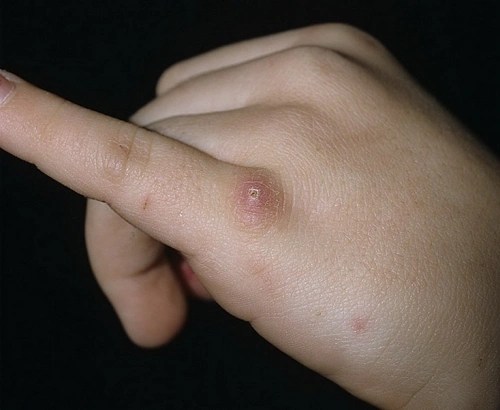
It is a purulent inflammation of the lymph nodes that occurs after a cat bite or scratch (the etiological factor is chlamydia, Rochalimaea henselae and Alipia GeH5). The incubation period lasts from 3 to 20 days. The disease is characterized by slow healing of injuries, regional lymphadenitis, and a febrile state. In the case of a typical form of the disease, at the site of an already healed wound after a bite or scratch, a small painful papule from 2 to 5 mm in diameter appears with a rim of skin hyperemia, which turns into a vesicle or pustule, and later into a small ulcer (not always), covered dry crust. After 2 weeks, regional lymph nodes increase to 5-10 cm in diameter, they are mostly painless. The axillary lymph nodes are more often enlarged, less often the cervical and inguinal lymph nodes. After 8 weeks they return to their original state. In 30% of children they melt.
Herpetic infection
Causative agent: herpes simplex virus,
The child is contagious as long as new elements appear.
A rash appears on the lips, skin, and oral mucosa (aphthous stomatitis) in the form of blisters with cloudy contents. During the rash period, there may be a high temperature.
Enteroviral vesicular stomatitis(Hand, foot, mouth syndrome) 
Pathogen: Coxsackie enterovirus A16,
Incubation period: 3-6 days. The child is contagious until the 10th day of illness. Temperature for 1-3 days. Bubbles appear on the mucous membrane of the mouth, palms, and soles, surrounded by a red halo, and go away on their own within 7-10 days.
infectious mononucleosis 
The causative agent is Epstein-Barr virus
Transmitted through close contact (for example, kissing).
Characterized by high fever for up to 10 days, sore throat, swollen lymph nodes, and nasal voice. The rash occurs when amoxicillin drugs (flemoxin, amoxiclav) are prescribed.
pseudotuberculosis And yersiniosis

Pathogen: Yersinia, incubation period 3-18 days.
It is transmitted by eating raw vegetables or through unboiled goat's milk.
Description: Usually there is a high temperature, there may be pain in the abdomen, joints, and diarrhea. Rash of various locations and shapes, typically of the “socks” and “gloves” type. The skin peels and comes off.
Scabies 
It is caused by a mite that makes microscopic passages in the thin skin of the interdigital spaces, wrists, stomach, genitals and other parts of the body. Severe itching of the skin occurs in the affected areas. Scabies is an extremely contagious disease and requires treatment by a dermatologist. It is transmitted from a person through close contact, through shared things. With scabies, the rash is accompanied by painful itching and looks like dotted elements, often located in pairs, 2-3 mm from each other. Often there is a layer of secondary infection (streptoderma).
Molluscum contagiosum 
Causative agent: poxvirus,
It is transmitted through close contact, through a shared bath, or swimming in stagnant bodies of water. Description: A rash up to 0.5 cm in diameter, with a “umbilical” depression in the center, a pearly tint, and when crushed, a cheesy discharge is released.
Bite marks
Bedbugs.
Representatives of the species Cimex lectularius reach sizes of 3-5 mm, they are active at night and feed only once a week. They usually live in floor cracks, furniture upholstery, and picture frames. The classic clinical sign of bedbug bites is a series of linear, itchy, urticarial papules that appear at night on exposed areas of the body. When examined using diascopy (by pressing a glass slide or spatula against the skin), a hemorrhagic point can be seen in the center of the rash. An examination of the bed linen, on which droplets of blood can be detected, will help make a diagnosis.
Fleas.
Fleas are minimally specific to their host, so human fleas can attack animals and vice versa. Human flea, Pulex irritans. They also bite on areas of the body covered by clothing. Flea bites present as urticarial lesions with small blue-red hemorrhages (purpura pulicosa). They are usually randomly located on the body. In children, lesions are sometimes papulovesicular and difficult to distinguish from childhood pruritus.
Hymenoptera.
This order includes bees, bumblebees, wasps and hornets. They sting with a special apparatus located at the back of the body, which is connected to a sac containing poison. Bee stings can often be seen on the feet of children who have walked barefoot across a meadow or lawn. The bite site should be carefully examined, as the sting may still be there. In this case, the sting must be carefully removed with small tweezers, being careful not to touch the sac with the poison. Wasps are more likely to sting children on the head, neck and arms because they are often attracted by the smell of food and drinks and because of this they “conflict” with people. Sometimes a wasp can fly into a glass and accidentally end up with its contents in a person’s stomach.
The local reaction to bites is well known to everyone - pain, erythema, swelling and, in some cases, blistering. This chain of events in the oral cavity can lead to obstruction (swelling and obstruction). In addition, systemic reactions may occur over the next few minutes, leading to pruritus, urticaria, anaphylaxis, and acute circulatory collapse in allergy sufferers.
Diptera.
This order includes flies and mosquitoes. Mosquitoes are most active in the early morning and evening. They bite exposed areas of the body. Mosquitoes are especially common near small bodies of standing water, as these are their favorite breeding grounds.
The mosquito bite initially presents as a pruritic, erythematous wheal, which then develops into a firm papule that persists for hours to days. Sometimes a blister or more severe local reaction with erythema, warmth and swelling develops at the site of the bite, usually on the extremities. Secondary impetiginization typically occurs as a result of scratching. Most often, this rash is accompanied by itching, but not very severe. The child's general condition does not suffer. He behaves as usual - plays, runs, throws things around, watches cartoons and eats with appetite.
2. ALLERGIC rash

Occurs after ingestion or contact with any allergen. Allergic rashes can be caused by environmental or food allergens. There are many allergens, but often they cannot be identified, even with every effort.
The most common allergens are house dust, animal hair, plant pollen, food, laundry detergents, especially at low water temperatures, natural wool, and some metals (for example, nickel on buttons, zippers, locks, buckles).
Food allergies can be caused by preservatives, dyes, chocolate, crustaceans, fish, eggs, strawberries, nuts, and tomatoes. Generally speaking, any food product can be an allergen, except perhaps table salt. It is also possible to be allergic to medications, often to penicillin antibiotics, etc.
An important sign that distinguishes allergies from infectious rashes is the good general condition of the child. The child may be irritable due to itching, but not drowsy, there is no loss of appetite or increase in body temperature.
If the rash is accompanied by swelling (especially on the face around the lips and eyes), be very careful and see a doctor right away. This may be a sign of a serious complication - Quincke's edema or even allergic shock. The spread of swelling to the area of the tongue and upper respiratory tract leads to suffocation. This condition requires emergency treatment in a hospital, sometimes even in an intensive care unit. Allergic reactions can occur even after lightly touching something. A classic example of this kind is rashes resulting from nettle or jellyfish stings.
By carefully assessing your child's diet and environment, you can probably figure out the cause of the allergy. Do not forget that mosquito bites in children also cause a local allergic reaction - as a result, multiple marks from mosquito bites can sometimes be mistaken for a rash.
It almost always appears suddenly, often accompanied by a profuse runny nose and lacrimation, and is very itchy. The rashes are raised and clearly visible. Even if there is no rash, the skin is irritated, red, and swollen. Taking antiallergic drugs eliminates both itching and the rash itself.
An allergic reaction appears quite quickly. Red spots of irregular shape, prone to merging and accompanied by severe itching, appear on the skin of the entire body or individual areas (cheeks, buttocks, behind the ears). The child's general condition may change: he may be lethargic or, conversely, too excited. Sometimes there is vomiting or loose stools. But more often the child feels well, but is very itchy. How can you help your child in this situation? First of all, it is necessary to exclude from his diet foods that cause an allergic reaction, even if they are very tasty and he loves them very much. Then you need to give the child sorbents - drugs that will remove the allergen from the child’s body. These include activated carbon, smecta, zosterin-ultra, filtrum. It is mandatory to take antiallergic drugs (the same suprastin or other drugs). Fenistil-gel and moisturizer are applied to the skin. It would be a good idea to see a pediatrician or dermatologist.
An allergic reaction can also occur when the skin comes into contact with some substances, such as washing powder, fabric softener, etc. In this case, the rash appears only in those areas that were in direct contact with the allergen. The tactics of parental behavior in this case are similar to those for food allergies. Additionally, the substance that caused the reaction should be removed from the skin - rinse off under running water. If you suspect the rash is caused by contact with clothing. Remember that in addition to unsuitable material, allergies can be caused by residue from washing powder or fabric softener. Try changing the manufacturer or using hypoallergenic hygiene products.
3. Rash when DISEASES OF BLOOD AND VESSELS is usually hemorrhagic in nature, i.e. occurs as a result of hemorrhages in the skin. Depending on the pathology, these can be large bruises of all colors of the rainbow, or a pinpoint rash covering the entire surface of the body.
Reasons: 1) A decrease in the number or dysfunction of special blood cells - platelets, which are actively involved in the blood clotting process (often congenital). 2) Violation of vascular permeability. 
In most cases, the rash is not palpable, except for inflammation of the walls of blood vessels. A hemorrhagic rash differs from other similar rashes in that when pressed it does not turn pale and does not disappear. The appearance of the rash is determined by the reasons for its appearance; for various diseases, it can have different sizes and colors. The color of hemorrhagic spots immediately after their appearance is red, then successively changes to blue, green, yellow, light brown, dark brown, dirty gray; The color disappears completely after 2-3 weeks.
Depending on the size and shape of the spots, petechiae (point hemorrhages), purpura (hemorrhages with a diameter of up to 1-2 cm), ecchymosis (hemorrhages with a diameter of more than 2 cm), and linear hemorrhages are distinguished.
The most common is a hemorrhagic rash on the legs, which can make diagnosis difficult, since such localization is typical for many diseases.
The cause of hemorrhagic rash can be hereditary and infectious diseases, steroid use, as well as various disorders that affect the blood vessels. A common cause of hemorrhagic rash in children under 5 years of age is an acute form of hemorrhagic vasculitis, a microvascular disease. Hemorrhagic vasculitis is most often accompanied by a hemorrhagic rash on the legs. Treatment is prescribed depending on the severity and form of the disease. As a rule, children are monitored at a dispensary during treatment. With proper and timely treatment, the disease has a favorable outcome.
Also, when a hemorrhagic rash appears in children, it is necessary to exclude hereditary diseases such as hemophilia and von Willebrand disease. Hemophilia is characterized by the appearance of subcutaneous hematomas, and any injuries are accompanied by extensive internal and external bleeding. Mostly men are affected by hemophilia. Von Willebrand disease leads to increased capillary fragility, which causes hemorrhage.
Such severe diseases as amyloidosis, Wegener's granulomatosis, thrombocytopenic purpura are accompanied by various types of hemorrhagic rashes and require immediate treatment.
Hemosiderosis of the skin is also accompanied by the appearance of a rash, which over time changes color from red to yellow or brown.
If a hemorrhagic rash appears, you should immediately consult a doctor and limit your mobility until diagnosis and hospitalization. In many cases, first aid is required in the very first hours after the rash appears, so there is no point in wasting time trying to self-treat. When a hemorrhagic rash appears in children, special care should be taken; even if they feel normal, they must remain in bed until the doctor arrives.
4. Due to the characteristics of children’s skin and frequent HYGIENE DEFECTS common diseases in infancy are prickly heat, diaper dermatitis, diaper rash. 
Avoid overly wrapping your baby. Try not to leave your baby in wet diapers or diapers. Bathe and wash your child more often, and also let his skin breathe - practice regular air baths.
Vesiculopustulosis- more unpleasant. 
This is a purulent inflammation of the mouth of the sweat glands in infants and young children, caused by pathogenic staphylococcus. It is characterized by pustular rashes, small blisters of white or yellowish color, which also affect infants. This is a fairly serious reason to worry and immediately seek medical help.
Bubbles appear on the back, chest, neck, legs and arms, even on the head. Then they burst, leaving crusts on the skin. It is dangerous because from the bursting blisters the infectious agent enters neighboring areas of the skin and “spreads” further throughout the body.
Having found such an abscess, carefully remove it with a cotton swab and alcohol and cauterize it with a strong (5%, dark, almost black) solution of potassium permanganate or a solution of brilliant green.
You will have to “paint” the child to prevent the infection from spreading. Wipe the skin around the pustules with alcohol, but only very carefully so as not to touch the pustule.
If you have vesiculopustulosis, you do not need to bathe your baby, as microbes from the bubbles enter the water and very quickly infect the entire skin.
What can you do
If you find your child has a skin rash, try to follow these rules:
1) It is always necessary to call a doctor at home, so that if you have an infectious disease, you do not infect others in the clinic and in transport. In addition, anyone with a rash should be isolated from pregnant women until a doctor says it is not rubella.
2) If you suspect your child has a meningococcal infection, or see any hemorrhagic rash, immediately call an ambulance
3) Before the doctor arrives, there is no need to lubricate the elements of the rash, especially with solutions containing dyes (for example, brilliant green). As you already understand, the main causes of rashes are internal. Consequently, you will not achieve a pronounced positive effect from lubricating the elements of the rash. However, it will be much more difficult for a doctor to make a diagnosis.


