Human lumbar muscles. Anatomy of the deep and superficial muscles of the human back. Deep muscles of the back and neck
Back muscles are numerous, as a result of which two groups are distinguished - superficial (two layers) and deep. The superficial muscles are attached to the shoulder girdle, shoulder and ribs, and skull. Deep muscles retained a primitive structure, forming a strong muscle mass, united by the common name “rectifier torso,” which stretches from the head to the pelvis. Innervated by branches of the brachial plexus.
|
Muscle name |
Attachment |
Muscle function |
Blood supply |
Innervation |
|||
Superficial back muscles: |
|||||||
|
1st layer of muscles: |
|||||||
|
1. Trapezius muscle (m. trapezius) |
external occipital protuberance, superior nuchal line, spinous processes of all thoracic vertebrae |
acromion process of clavicle, acromion, spine of scapula |
the upper beams – raise the scapula, the lower ones – lower it, when fixing shoulder girdle– pull the head back, with unilateral contraction – tilts the head in the appropriate direction, and turns the face in the opposite direction |
occipital artery, intercostal arteries, transverse cervical artery |
accessory nerve |
||
|
2. Latissimus muscle back (m.latissimusdorsi) |
spinous processes of 6 lower thoracic and all lumbar vertebrae, dorsal surface of the sacrum, iliac crest, 9-12 ribs |
crest of the lesser tubercle of the humerus |
brings the shoulder to the body and pulls the upper limb back to the midline, rotating it inward; with a fixed limb, it brings the body closer, |
subscapular artery |
subscapular nerve |
||
|
2nd layer of muscles: |
|||||||
|
1. Muscle that lifts the scapulae (m. levatorscapulae) |
transverse processes of the 4 upper cervical vertebrae |
upper angle of the scapula |
raises the scapula, especially its inner corner, with the scapula strengthened, tilts the neck backwards and its side |
transverse cervical artery intercostal arteries |
dorsal scapular nerve dorsal scapular nerve |
||
|
2. Rhomboid muscles: |
spinous processes of 4-5 upper thoracic vertebrae; |
medial edge of the scapula below the spine; |
|||||
|
Large (m. rhomboideusmajor) |
spinous processes of the 2 lower cervical vertebrae |
medial edge of the scapula above the spine |
pull the scapula to the spinal column and the top, press the scapula to chest |
||||
|
Small (m. rhomboideus minor) |
|||||||
|
3. Rear upper serratus muscle(m. serratus posterior superior) |
spinous processes of the 2 lower cervical and 2 upper thoracic vertebrae |
raise the upper ribs, participating in the act of inhalation |
transverse cervical artery, intercostal arteries |
intercostal nerves |
|||
|
4. Posterior lower serratus muscle (m. serratus posterior inferior) |
spinous processes of the 2 lower thoracic and 2 upper lumbar vertebrae |
lower 9-12 ribs, participating in the act of exhalation |
lumbar artery |
dorsal branches of the spinal nerves |
|||
Deep back muscles |
|||||||
|
1. Sacrospinalis muscle or erector spinae muscle. It has 3 parts: |
lateral ridge of the sacrum |
transverse processes of 4-6 cervical vertebrae, angles of the upper 5-7 and lower 8-9 ribs |
with bilateral contraction it is a powerful extensor of the spinal column, holds the torso in an upright position, with unilateral contraction it tilts the spinal column in the corresponding direction; the upper tufts pull the head in their direction |
dorsal branches of the intercostal arteries and vertebral artery |
dorsal branches of the spinal nerves |
||
|
Iliocostal muscle (m. iliocostalis) |
dorsal surface of the sacrum, transverse processes of the lumbar and lower 6-7 thoracic vertebrae |
angles of the 10 lower ribs, mastoid process, transverse processes of all thoracic vertebrae |
|||||
|
Longissimus dorsi (m. longissimus) Rating 4.21 | |||||||
Without them, none of his movement would have been possible. At the same time, they are used by the human body not only for movements, but also play an important static supporting role, supporting and fixing among themselves both individual elements of the spine - the vertebrae, and the spine as a whole, setting and maintaining its natural curves. Thus, the back muscles do not rest for a minute in an active, awake person in an upright position. One can imagine how tediously hard their work is.
Main functions of the back muscle
Let us systematize the above by listing the main functions performed by the back muscles:
- Permanent fixation of the vertebrae among themselves to provide immovable support. Without this, none of our turns and bends, flexions and extensions would be possible.
- Keeping your back and neck upright
- Ensuring motor activity of all spinal regions
- Maintaining the natural curvature of the back (cervical and lumbar lordosis and thoracic kyphosis) within the required limits
- Cushioning of the spine during active movements leading to shocks, shocks and vibrations
The back muscles are not a homogeneous muscle mass. There are many of them and they different sizes, types and functions. They are superficial and deep. Both types consist of two layers.
Let's look at the back muscles from an anatomical perspective and highlight the most important of them.
The most important of them, supporting the spine vertically, is erector muscle (erector):
This is a powerful muscle located on the surface of the deep ones, running along the entire spinal column and filling all the depressions from the spinous processes to the costal surfaces.
All back muscles have their own characteristics, the main ones being:
- Start
- fastening
- functions performed by it
So, the characteristics of the erector dorsi muscle.
Start:
- posterior surface of the sacrum
- posterior iliac crest
- spinous processes of the lower lumbar vertebrae
- thoracolumbar fascia
- lateral - iliocostal
- medially - spinous
- between these two is the longest
All parts have different places fastenings:
- ribs
- transverse and spinous processes
Functions:
- vertical position support
- extension of the spine and its inclinations
- head turns
- partially involved in respiration
On the surface of the back you can see two more of the largest intersecting muscles:
These are the trapezius and latissimus. Their name itself speaks volumes about their external characteristics.
Trapezius muscle:
Two muscle triangles on either side of the spine, with a base near the vertebral axis and an apex near the acromion of the scapula, form a trapezoid shape. Their tendons are short, and only at the border of the cervical and thoracic regions are they slightly longer, forming a diamond-shaped area.
Start:
- spinous processes of the thoracic vertebrae
- nuchal ligament - a thin elastic triangular plate coming from the spinous processes and attached to the occipital crest and protrusion
Fastening:
- Acromial end of the clavicle
- Acromion and scapular axis
Functions:
- Hands up
- Movement of the shoulder blades - raising, lowering, bringing the shoulder blades closer together
- Head tilts in different directions
Latissimus
The latissimus dorsi muscle covers a wide area: from the humerus to the iliacus, crossing the back obliquely, occupying its entire bottom part and forming the lumbar triangle below.
Covered at the top in a small area near the trapezoidal spine.

Start:
- spinous processes of the extreme 4 - 6 vertebrae of the thoracic region and all vertebrae of the lumbar and sacral region
- four lower ribs to which muscle teeth are attached
- thoracolumbar fascia - a membrane within which there are deep muscles of the lumbar and thoracic. Attachment occurs by fusion of the smooth tendon area (aponeurosis) of the latissimus muscle with the fascia
- iliac crest
Place of its attachment:
- crest of the lesser tubercle of the humerus
The latissimus dorsi muscle performs the following functions:
- Provides movement of the shoulder with pulling the arm back to the spinal axis with pronation(rotating inward)
- Allows the body to be pulled towards the arm while securing the latter. With its powerful development it becomes possible brachiation- movement with the help of hands, which is always readily used by our relatives - monkeys
Rhomboid major muscle
It is located under the trapezoid and has the shape of a rhombus.
Start
- two outermost cervical vertebrae
- 4 upper pectorals
Fastening
- to the medial edge of the scapula
Functions:
- Attraction of the scapula towards the spine and upward
- Fixation of the medial edge of the scapula to the chest
Big round
The teres major muscle of the back has a flat, elongated shape. Covered at the back latissimus dorsi, in front - the triceps humerus.
Start:
- inferior angle of the scapula
- fascia of the infraspinatus (filling the scapula fossa) muscle
Fastening:
- crest of the tubercle of the humerus with the help of tendons located in the subtendinous bursa
It belongs to the muscles of the upper limb, hence its importance in performing such arm movements:
- pulling the arm down and back
- adduction- bringing the arm to the body
- pronation- inward rotation
The role of the oblique muscle in the functionality of the spine. In the muscles of the lower torso you can see another important object, which, although it is an abdominal muscle, in its functions plays a big role for the lower back. This is the oblique abdominal muscle.
There are two types of it:
External oblique
Start:
- outer surface 5 - 12 ribs
Fastening:
- external line of iliac crest
- pubic symphysis
- Linea alba - white collagen fibers located along the midline of the abdominal wall
Internal oblique- this is a wide muscle-tendon plate located under the external muscle
Start:
- intermediate line of the iliac crest
- thoracolumbar fascia
- lateral (side) part of the inguinal ligament
Fastening:
- outer cartilaginous surface of the lower ribs
- linea alba (here the attachment is carried out using a wide tendon)
The external oblique and internal oblique are involved in a number of functions:
- Movements of the chest and pelvis
- Torso rotations
- Spinal flexion
From this it can be seen that in the motor function of the spine, not only the muscular system of the back, but also the abdomen plays a large role.
Two weak points in the back. The external oblique and internal oblique muscles are involved in the formation of two important areas of the human body, weakness of which can contribute to the development lumbar hernia. These are the so-called:
Lumbar triangle, also called Petit's triangle
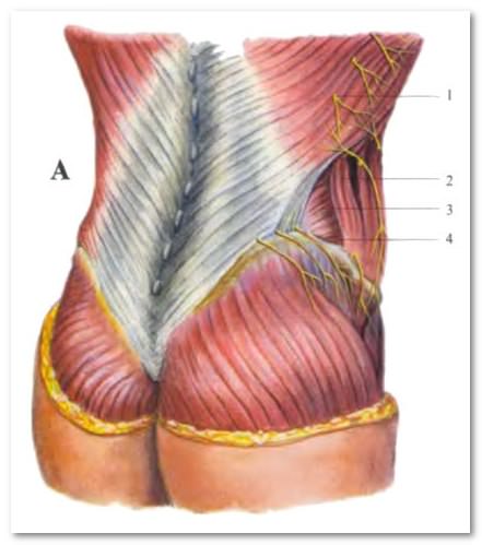
This is a section of the rear wall, limited on three sides:
- behind - latissimus muscle
- in front - external oblique
- below - the iliac crest
The bottom of the triangle is formed by an oblique and transverse muscle belly. Essentially, a triangle is a small gap between the edges of the latissimus and external oblique, which does not occur in everyone, but in about 75% of people
Lumbar quadrangle of Grunfeld-Lesgaft
Bounded on four sides:
- superior lateral – 12th rib and serratus inferior muscle
- lateral - external oblique (its posterior edge)
- bottom - edge of the internal oblique
- medial - edge of the sacrospinous
This place is considered weak due to the fact that the oblique abdominal muscles do not cover the quadrangle, so it is not strengthened, and in this place there is a high risk of developing a lumbar hernia.
Taking care of the back muscular system
Anatomy of the back muscles and description muscle functions show the vital need for their strengthening. Often incorrect posture, which then leads to many diseases of the spine, occurs due to muscle asymmetry, which can only be eliminated by intensive training. In addition to regular back exercises, special ones are also performed using exercise machines.
In order to muscular system your back was in good shape, try to follow three simple rules:
- Be sure to start your day with morning exercises
- Stay less in a relaxed “jelly” position
- Sleep on a hard, flat surface or on a special orthopedic mattress
But it would be wrong to constantly punish our muscles, keeping them in a “black” body and not allowing us to relax even for a minute. So be sure to relax them. Relaxation methods:
- Exercises for relaxation.
They must be carried out in the intervals between periods of muscle activity. - Relaxing massage
Especially recommended for athletes after training and people doing physical work after the end of the working day. - Water procedures: pine baths, back swimming
Protect your back muscles from unnecessary strain and hypothermia. This may lead to
Muscles of the body: back, chest, abdomen, attachment points, functions. Weak spots on the anterior abdominal wall: linea alba, inguinal canal, umbilical ring.
Main questions:
1. Classification of back muscles: superficial and deep back muscles.
2. Back muscles: groups, topography, points of origin and insertion, functions.
3. Classification of chest and abdominal muscles.
4. Chest muscles: groups, topography, points of origin and insertion, functions.
5. Diaphragm: parts, holes, meaning, change in shape during breathing. Weak spots.
6. Abdominal muscles: groups, topography, points of origin and insertion, functions.
7. Abdominal Press. Weak spots on the anterior abdominal wall: linea alba, umbilical ring, inguinal canal, inguinal ligament.
The muscles of the trunk are divided into muscles of the back, chest and abdomen.
Back muscles.
The back muscles are paired, divided into deep and superficial. Greatest development from the back muscles reach the muscles of the superficial layer, belonging to the type strong muscles, performing mainly static work. They extend throughout the back and back of the neck from the sacrum to the occipital bone. The places of origin and attachment of these muscles occupy large surfaces and therefore, when contracting, the muscles develop great force, holding the spine in a vertical position, which serves as a support for the head, ribs, entrails and upper limbs. General principle The location of the deep back muscles is the formation of several layers, and the deeper the muscle is located, the shorter it is.
Superficial back muscles.
Trapezius muscle– flat, triangular in shape, located on the upper back and back of the neck. It starts from the occipital bone, nuchal ligament, supraspinous ligament and spinous processes of the 7th cervical and all thoracic vertebrae. Attached to the acromial part of the clavicle, the humeral process and the spine of the scapula. Function: when the upper fascicles contract, the scapula rises; middle beams bring the scapula closer to the spine; the lower ones lower the scapula. With bilateral muscle contraction, the shoulder blades move closer to the midline, and with fixed shoulder blades, the head and neck tilt backward.
Latissimus dorsi muscle– flat, the widest of all the muscles of the body, triangular. It lies superficially in the lower parts of the back and in the posterolateral parts of the chest. It starts from the spinous processes of the six lower thoracic and all lumbar vertebrae, the sacrum, the ilium, 9-10 ribs, and the thoracolumbar fascia. Attached to the crest of the lesser tubercle of the humerus. When contracted, the muscle pulls the arm back, turns it inward, and takes part in breathing movements.
Levator scapulae muscle– has a flat, slightly rounded shape, located on the lateral surface of the neck, covered in the upper parts in front by the sternocleidomastoid muscle, and in the lower parts in the back - by the trapezius. The muscle begins with four tendon teeth from the transverse processes of the first four cervical vertebrae and, moving from top to bottom and posteriorly, is attached to the medial edge of the scapula at the upper internal angle. Function: the muscle lifts the inner angle of the scapula upward and lower - turns inward. With a fixed shoulder blade, it tilts the cervical part of the spine in its direction and rotates it somewhat.
Rhomboid major and minor muscles– located under the trapezius muscle, often fused to form one muscle. The muscles begin on the nuchal ligament in the region of the spinous process of the 2-5 and 7 cervical vertebrae, the first thoracic vertebra and, moving obliquely downwards, attaches to the medial edge of the scapula. Function: brings the scapula closer to the spine, while simultaneously moving it upward.
Serratus posterior superior– located in the upper back under the rhomboid muscle. The tendinous part of the muscle starts from the lower part of the nuchal ligament, the spinous process of the seventh cervical and two upper thoracic vertebrae and passes into the muscular part. The latter ends with four teeth near the corners of the second to fifth ribs. Function: the muscle raises the upper ribs, participating in inhalation; unilaterally - tilts the spinal column to the side, bilaterally - extends the spinal column.
Serratus posterior inferior muscle- lies in the lower back under the latissimus muscle, originates from the spinous processes of two or three lower thoracic and two upper lumbar vertebrae and, turning into four wide teeth, is attached to the lower edges of the last four ribs. Function: pulls the lower ribs down, participating in the act of exhalation.
They are attached to the skeleton of the shoulder girdle and to the humerus and are located in two layers. The first layer consists of the trapezius muscle and the latissimus dorsi muscle, the second layer consists of the rhomboid major and minor muscles and the levator scapulae muscle.
Trapezius muscle- flat, triangular in shape, with a wide base facing the posterior midline, occupies the upper and posterior region of the neck. It begins with short tendon bundles from the external occipital protrusion, the medial third of the superior nuchal line of the nuchal bone, from the nuchal ligament, the spinous processes of the 7th cervical vertebra and all thoracic vertebrae, and from the supraspinous ligament. From the origin, the muscle bundles are directed, noticeably converging, in the lateral direction and attached to the bones of the shoulder girdle. The superior muscle bundles pass downwards and laterally, ending on the posterior surface of the outer third of the clavicle. The middle bundles are oriented horizontally, extend outward from the spinous processes of the vertebrae and are attached to the acromion and scapular spine. The lower muscle bundles follow upward and laterally, passing into the tendon plate, which is attached to the scapular spine. Tendon origin trapezius muscle It is more pronounced at the level of the lower border of the neck, where the muscle is widest. At the level of the spinous process of the 7th cervical vertebra, the muscles of both sides form a well-defined tendon area, which is found in the form of a depression in a living person.
The trapezius muscle is located superficially throughout its entire length, its upper lateral edge forms the posterior side of the lateral triangle of the neck. The lower lateral border of the trapezius muscle crosses the latissimus dorsi muscle and the medial border of the scapula externally, forming the medial border of the so-called auscultation triangle. The lower border of the latter runs along the upper edge of the latissimus dorsi muscle, and the lateral border runs along the lower edge of the rhomboid major muscle (the dimensions of the triangle increase when bent forward in shoulder joint hand when the scapula moves laterally and anteriorly).
Function: simultaneous contraction of all parts of the trapezius muscle with a fixed spine brings the scapula closer to the spine; the upper muscle bundles raise the scapula; the upper and lower bundles, while simultaneously contracting, forming a pair of forces, rotate the scapula around the sagittal axis: the lower angle of the scapula moves forward and in the lateral direction, and the lateral angle moves upward and medially. With a strengthened scapula and contraction on both sides, the muscle extends the cervical spine and tilts the head back; with unilateral contraction, it slightly turns the face in the opposite direction.
Latissimus dorsi muscle- flat, triangular in shape, occupies the lower half of the back on the corresponding side.
The muscle lies superficially, with the exception of the upper edge, which is hidden under the lower part of the trapezius muscle. Inferiorly, the lateral border of the latissimus dorsi muscle forms the medial side lumbar triangle(the lateral side of this triangle is formed by the edge of the external oblique muscle of the abdomen, the lower - by the iliac crest. It begins with an aponeurosis from the spinous processes of the lower six thoracic and all lumbar vertebrae (together with the superficial plate of the thoracolumbar fascia), from the iliac crest and the median sacral crest. Muscle bundles follow upward and laterally, converging towards the lower border axillary fossa. At the top, muscle bundles are attached to the muscle, which start from the lower three to four ribs (they extend between the teeth of the external oblique abdominal muscle) and from the lower angle of the scapula. Covering the lower corner of the scapula from behind with its lower bundles, the latissimus dorsi muscle sharply narrows and spirals around the teres major muscle. At the posterior edge of the axillary fossa it passes into a flat thick tendon, which is attached to the crest of the lesser tubercle of the humerus. Near the place of attachment, the muscle covers from behind the vessels and nerves located in the axillary fossa. It is separated from the teres major muscle by a synovial bursa.
Function: brings the arm to the body and turns it inward (pronation), extends the shoulder; lowers the raised hand; if the arms are fixed (on the horizontal bar), the torso is pulled towards them (when climbing, swimming).
Levator scapulae muscle begins with tendon bundles from the posterior tubercles of the transverse processes of the upper three or four cervical vertebrae (between the places of attachment of the middle scalene muscle - in the front and the splenius muscle of the neck - in the back). Moving downward, the muscle attaches to the medial edge of the scapula, between its upper angle and the spine of the scapula. In its upper third the muscle is covered by the sternocleidomastoid muscle, and in the lower third by the trapezius muscle. Immediately anterior to the levator scapulae muscle are the nerve to the rhomboid muscle and the deep branch of the transverse cervical artery.
Function: raises the scapula, simultaneously bringing it closer to the spine; with a strengthened shoulder blade, it tilts the cervical part of the spine in its direction.
Rhomboid minor and major muscles often fuse and form one muscle.
The rhomboid minor muscle starts from the lower part of the nuchal ligament, the spinous processes of the 7th cervical and 1st thoracic vertebrae and from the supraspinous ligament. Its bundles pass obliquely - from top to bottom and laterally and are attached to the medial edge of the scapula, above the level of the spine of the scapula.
The rhomboid major muscle originates from the spinous processes of the 2-5 thoracic vertebrae; attaches to the medial edge of the scapula - from the level of the spine of the scapula to its lower angle.
The rhomboid muscles, located deeper than the trapezius muscle, themselves cover the posterior superior serratus muscle and partially the erector spinae muscle.
Function: brings the scapula closer to the spine, while simultaneously moving it upward.
Two thin flat muscles are attached to the ribs - upper and lower rear serrations.
The superior posterior serratus muscle is located in front of the rhomboid muscles, begins in the form of a flat tendon plate from the lower part of the nuchal ligament and the spinous processes of the 6-7 cervical and 1-2 thoracic vertebrae. Directing obliquely from top to bottom and laterally, it is attached with separate teeth to the posterior surface of 2-5 ribs, outward from their corners.
The back muscles are the largest group of fibers that form the muscular corset of the human spine. Anatomical structure quite complex and due to the need to perform numerous functions.
General Anatomy and Functions
The surface of the back is divided into several anatomical zones:
- Vertebral (central). This area contains the muscles that straighten the trunk.
- Scapular. The muscles located here allow you to lift your upper limbs.
- Subscapular. The work of this zone allows you to bend the body to the sides.
- Lumbar. Carries the main axial load.
- Sacral.
In these areas, the muscles are located in 2 layers: the so-called superficial and deep back muscles. Coordinated work of all layers muscle fibers allows you to perform the following functions:
- Maintaining your back in an upright position.
- Formation of physiological curves of the spinal column (kyphosis and lordosis).
- Turn the body in different directions.
- Tilts forward, backward, left, right.
- Movement of the upper limbs.
- Participating in inhalation and exhalation by raising and lowering the ribs.
- Participation in head movement (turning, bending, throwing back).
- Implementation of rotation (rotation) of the body.
- Creating a protective layer for joints, internal organs and bone structures.
- Participation in the absorption of excessive loads on the spine.
Surface layer
The superficial back muscles are attached at one end to the spinous processes of the vertebrae, and at the other to various bone structures of the upper part of the body (scapula, collarbone, ribs, humerus). If this muscle group is sufficiently developed, the back acquires a beautiful relief, so in fitness and bodybuilding much attention is paid to pumping up this particular area.
Trapezoidal
A wide and flat muscle, shaped like a trapezoid, consisting of two triangles, with their base facing the spinal column and their apex facing the process of the scapula. With unilateral contraction, the trapezius tilts the head to the side, and with bilateral contraction, it throws it back, straightening the cervical spine.
Latissimus
Located in the lower back. The latissimus muscle occupies a superficial position, with the exception of the uppermost section, which is covered by the trapezius. Main functions:
- Controls the movement of the shoulder (brings it to the body, pulls it back, rotates).
- Pulls the torso up when the arms are fixed (for example, on a horizontal bar).
- Participates in the act of breathing, displacing the lower ribs.
The latissimus muscle is sometimes called the wings because in bodybuilders it protrudes at the level of the armpits, giving the body a V-shaped appearance.
Big round
The teres major muscle of the back is located immediately below the latissimus, forming back axillary fossa and performing the same function: provides shoulder movement. Despite its name, it is a small teres major muscle and occupies a very small area.
Large and small diamond-shaped
They are located in the second layer of the superficial muscle layer. These spinal muscles are attached with one edge to the spinous processes of the vertebrae (the small one to the sixth and seventh cervical vertebrae, and the large muscle to the first five thoracic vertebrae). These muscle fibers control the movement of the shoulder blades: they bring the shoulder blades closer to the chest, pull them towards the spine and up, and play an important role in the formation of an upright posture. When the rhomboid muscles are weak, the shoulder blades move forward and a stooped position develops.
Upper and lower rear gears
These muscles are quite thin and invisible. With their lower edge they are attached to the ribs, taking part in respiratory movements. The superior serratus muscle helps to inhale, and the lower one helps to exhale. The innervation of these muscle fibers is carried out by intercostal nerves, pinching of which causes intercostal neuralgia, familiar to many people.

Deep muscles of the back and neck
This group is located between the two layers of deep fascia of the back and the spine, makes up the majority of the muscle mass and is critical for maintaining correct posture.
Belt
Despite the name, the splenius muscle belongs to the group of spinal muscles. Its longitudinal muscle fibers begin from the lower cervical and upper thoracic vertebrae. Unilateral tension of the splenius neck muscle causes the head to tilt in the corresponding direction, and bilateral tension leads to extension cervical region spine and head tilt.
Spinal erector
It is the most powerful and longest paravertebral muscle of the back, extending the spine and holding it in upright position. With unilateral tension in the muscle fibers, the torso tilts to the side. The upper muscle bundles pull the head in their direction, and the lower muscle fibers lower the ribs.
The erector spinae comes from the posterior surface pelvic bones and spinous processes of the lower lumbar vertebrae. Heading upward, it is divided into the iliocostal muscle, the medial spinous muscle and longissimus muscle backs.
Transverse spinous
These muscles of the spinal column are located along the entire spine and fill the space between the transverse processes of the underlying and spinous processes of the overlying vertebrae and are covered by the erector spinae muscle. The muscle bundles have different lengths, so the transverse spinalis muscle consists of three independent muscle groups: semispinalis, multifidus and rotator cuffs.
Interspinous
They connect the spinous processes of adjacent vertebrae and are short muscle bundles arranged in pairs. Topographically, the interspinous muscles of the neck, chest and lower back are distinguished. The main function is to participate in the extension of the spinal column.
Intertransverse
These muscles of the spinal column in the form of short muscle bundles connect the transverse processes of adjacent vertebrae. Topographically, the posterior/anterior intertransverse muscles of the neck are distinguished; intertransverse muscles of the chest, lateral and medial intertransverse muscles of the lower back. Function – they ensure that the spine is held in an upright position, and when there is tension on one side, the torso is tilted.
Quadratus lumborum muscle
This large muscle layer is located in the lower back and is formed by three bundles of fibers. The first group of muscle fibers is the largest. It is attached to the last (lower) rib and the posterosuperior part of the iliac crest. The second group consists of oblique fibers connecting the spinous processes of the first three lumbar vertebrae with the iliac crest and the iliopsoas ligament.
The third group of fibers stretches from the 12th rib to the processes of the lumbar vertebrae. With bilateral contraction of this muscle, it extends lumbar region the spine, and in case of one-sidedness, a tilt to the side is carried out. In addition, if the pelvis is fixed, then with unilateral contraction of the quadratus muscle, the torso rotates in the direction of contraction. This muscle is also involved in ensuring forced breathing, stabilizing the lower rib.
The quadratus muscles are such powerful lumbar stabilizers that if they are paralyzed, a person will not be able to walk.
Kinesiology of the back muscles
Kinesiology (from the Greek words “movement” and “knowledge”) is a practical science that studies muscle movement. There is hardly an adult who has not experienced back pain at least once. Most often, these pains are associated with excess tone (spasm) of certain muscle fibers caused by pathology of the spine or internal organs or lack of muscle mass.

Research in the field of kinesiology laid the foundation for kinesitherapy
Specialists (kinesiologists and kinesitherapists) identify the presence of muscle blocks in the back and select special complex exercises to eliminate tension. Thanks to this therapy, people not only experience pain relief, normal movement of the back and neck is restored, but also metabolism and general well-being improve, and the functioning of many internal organs is normalized.
Thus, the superficial and deep muscles of the human back are a powerful and balanced mechanism that provides various movements of the torso and upper shoulder girdle, as well as keeping the human body in an upright position.
The muscles of the back take part in ensuring the normal functioning of internal organs. Maintaining good tone and sufficient mass muscle corset by using physical exercise And proper nutrition is an important component of the health of the spine and the entire body as a whole.


