Vastus medialis muscle treatment. Where is the quadriceps femoris muscle located: photo of the origin and attachment of the muscle. Quadriceps femoris
The quadriceps femoris has the maximum volume in relation to the rest of the muscle mass. It received this name because it contains four large muscle elements. Despite its power, this muscle is more susceptible to damage than others because the fibers are close to the surface. Beginning athletes often suffer from muscle tears. This is why you need to know muscular anatomy, especially for those who decide to get in shape in the gym.
Anatomy of the quadriceps femoris muscle
The quadriceps is a kind of conglomerate of four muscle masses:
- direct;
- lateral;
- medial;
- intermediate.
In the patella of the femur, all these heads form a common tendon, its attachment goes to the superficial structures of the tibia and the kneecap.
The anatomy of the powerful quadriceps femoris muscle is based on the structure of its components.
The rectus femoris muscle arises from the acetabulum. Between the bone surface and muscle tissue is the joint capsule. Next, the muscle turns down the front side of the hip joint, protruding closer to the skin between the sartorius muscle element and the tensor fasciae lata. The end of the muscle flows into the tendon of the quadriceps femoris muscle, which is attached to the top of the kneecap. Basically, hip flexion is carried out with its help.
The largest muscle fiber of the quadriceps muscle is the vastus lateralis. Its top is attached by tendon bundles to the head of the femur and to the lateral intermuscular septum. Below it joins the tendon common to the quadriceps mass.
 The vastus intermedius muscle starts on the ventral part of the thigh bone. It attaches to the top of the knee cup and participates in the creation of a single tendon bundle.
The attachment of the upper part of another muscle mass, the broad medial one, occurs in the area between the two trochanters, near the medial lip of the linea aspera. Then it passes along the medial side of the thigh. At the bottom it also joins into a tendon with the rest of the quadriceps.
The blood supply to the quadriceps muscle is provided by the femoral artery, which is a continuation of the iliac artery. The innervation of muscle tissue is carried out by the femoral nerve, which regulates motor abilities.
To understand exactly where these hip muscles are located, you can look at the tense legs of athletes. In people who are keen on bodybuilding and powerlifting, the muscle mass is very pronounced.
Basic functions of the quadriceps
There are two important functions performed by the quadriceps femoris muscle: static and dynamic. Thanks to the first, a person is able to stay upright and maintain balance. The quadriceps holds the joint of the knee, that is, it does not allow it to move and the limbs to “give way.” Dynamic function refers to the ability of the knee joints to remain stable during intense movements.
 The quadriceps muscles take part in flexion and extension of the lower extremities, pulling them towards the body, and tilting the body in the femoral area. Its largest part, the lateral one, performs a damping function and is involved in straightening the knee joint. The appearance of the hips on the outside depends on the development and shape of this massif.
Functional features of the other three muscles, in addition to flexion and extension:
- The middle muscle prevents the kneecap from moving.
- The intermediate helps to straighten the limb at the knee joint when a person runs, jumps or squats.
- The rectus muscle covers the remaining fibers of the quadriceps. In addition to the protective function, it forms the roundness of the top of the leg.
The quadriceps has two types of muscle fibers - fast and slow. The latter help a person maintain balance, and the former prevail in those areas of the muscular elements that are responsible for elasticity.
Quadriceps workouts
The quadriceps bear the main static load while maintaining balance. The quadriceps muscle makes up seventy percent of the muscle mass of the limb, and its development is fundamental when training the legs. Working on the muscles that are part of it allows you to achieve bodily harmony, endurance, and increase physical strength. Active training of the lower extremities improves the functioning of the excretory and genital organs, accelerates blood flow, relieves congestion and relieves the hip and knee joints from unnecessary stress.
In gym
 The muscle mass is easily injured. Before classes, a warm-up is necessary. The beginning of the workout can be a five-minute jog on a special track, which helps to warm up the muscle mass of the legs. In addition to the treadmill, exercises with a skipping rope and simple squats are suitable. Also, as a warm-up, they goose-step around the hall.
Squats. When performing, there is a risk of achieving such muscle relief that the limbs become like the legs of an overweight person, and not an athlete. To prevent this from happening, it is necessary to take into account the ratio of the length of the torso and legs. With a long thigh, the lumbar muscles become overloaded.
Athletes who experiment with weights tend to overload the muscle fibers of the gluteal region, which is the wrong approach to training. Because of this, squats do not give the required result.
Do squats with a barbell placed on your chest. This method allows you to use the necessary muscle groups, which will make the training effective and protect against possible injury or pumping of the buttocks.
Squats in a hack machine. Here the lumbar region is almost not loaded, and the different position of the feet makes it possible to load the quadriceps. A hack trainer is a device for training the muscles of the lower body. It straightens your back and allows you to simulate squats with a barbell. The lower limbs are placed on a supporting platform approximately shoulder-width apart and bent at the knees at a right angle. The back rests firmly against the back of the training unit. They perform a squat and simultaneously move the platform with the load. This works the quadriceps, adductors, buttock muscles and tendons behind the knees.
 Leg press. With a small distance between the legs, the load is transferred to the quadriceps muscle. An extremely low position of the platform will not work, but this is not necessary. A significant part of the movements occurs at the top. Relaxation of the quadriceps in the lower position and full extension in the upper position are not allowed. The task is not to lift the weight, but to strive to achieve straightening of the legs.
Leg extensions. The lesson should be carried out alternately on each limb. In the lower position, you cannot sharply lower your legs, and at the top, hold the straightened limb for a long time. The optimal number of repetitions is approximately 10 to 15 times.
Lunges with dumbbells. The workout must be performed eight to twelve times in three sessions. It involves different muscle groups, but the quadriceps muscle is subjected to the greatest load.
At home
Squats with weights can be done not only in the gym, but also at home; you just need to choose the appropriate load and change it. Adjustable weight dumbbells are perfect for this exercise.
 What else can be included in the complex of home strength training:
- Lunge with stretch. With your feet shoulder-width apart, take a wide step forward. Return to the starting position. Without pausing, take a step with your other foot.
- Lunges to the side. The process is similar, only you need to step to the right and left.
- Sumo squats. Legs are spread wide, feet pointing outward. The squat is performed slowly, with tension in the quadriceps.
- Jumping in place according to the technique: legs together, then apart.
To increase the volume of muscle tissue and strength capabilities, classes are conducted at a slow pace. To burn subcutaneous fat, move faster. The training is carried out with a number of visits of at least twenty-five.
Execution Rules
Strength exercises for the quadriceps femoris muscle require adherence to a certain technique. It is not allowed to fully straighten the leg at the knee joint to avoid injury even when using light weight. You also need to forget about doing the exercise in jerks. If you are overweight, reduce it.
Control your breathing during exercise. Inhale while bending, exhale while extending.
 The quadriceps femoris muscle is the largest muscle, and loads on it cause changes in blood pressure. The muscle mass pumps a colossal amount of blood in the body, which is a possible reason for the increase in heart rate. Between approaches, you need to take a two-minute rest to normalize the pressure. If you feel dizzy after the lesson, the break will be extended. During the training process, drink water, otherwise you may become dehydrated and lose strength.
The quadriceps is a quadriceps muscle that contains both fast and slow muscle fibers. For the harmonious development of muscle mass in this area, you should not use only power loads. Be sure to include cardio training and static exercises.
Quadriceps femoris, m. quadriceps femoris(see Fig. , , , , , , , , , , , ). Each of the four heads has its own origin, but, approaching the knee area, they all pass into a common tendon, which covers the patella and attaches to the tibial tuberosity.
Rectus femoris muscle, m. rectus femoris(see figure), the longest of the four heads. Occupies the anterior surface of the thigh. It begins with a thin tendon from the inferior anterior iliac spine, the supraacetabular groove. Heading down, the muscle passes into a narrow tendon, which is part of the common tendon of the quadriceps femoris muscle. Having reached the tibia, the muscle tendon attaches to the iliac tuberosity. Below the patella, this tendon is called the patellar ligament, lig. patellae
Vastus medialis muscle, m. vastus medialis(see Fig.) occupies the anteromedial surface of the lower half of the thigh. The muscle bundles that form it are directed obliquely from top to bottom and from the inside to the front. The front is somewhat covered by the rectus muscle. The muscle originates from the medial lip of the linea aspera of the femur and, moving downwards, passes into the broad tendon, which is partially woven into the common tendon along with the rectus muscle, and partially attached to the medial edge of the patella, forming the medial suspensory ligament of the patella.
Vastus lateralis muscle, m. vastus lateralis, occupies almost the entire anterolateral surface of the thigh. From above it is somewhat covered by the muscle that strains the fascia lata, and in front by the rectus femoris muscle. The muscle bundles are directed from top to bottom and from the outside to the front (see Fig.).
The muscle originates from the greater trochanter, the intertrochanteric line and the lateral lip of the broad line of the thigh. Heading down, the muscle passes into the broad tendon, which is part of the common tendon of the quadriceps muscle and is involved in the formation of the lateral suspensory ligament of the patella.
Vastus intermedius muscle, m. vastus intermedius(see Fig.) is located on the front surface of the thigh between the vastus medialis and lateralis muscles, directly under the rectus femoris muscle. This muscle is the weakest among the other heads. It begins on the anterior surface of the femur - from the intertrochanteric line and, going down, passes (almost half its length) into the broad tendon, which in the distal part joins the tendon of the rectus femoris muscle, passing into the common tendon of the quadriceps muscle.
Thus, all four heads of the muscle that form the quadriceps femoris muscle pass into a tendon that includes the patella and attaches to the tibial tuberosity. Synovial bursae are located in front and behind the tendon (see Fig.):
- A) subcutaneous prepatellar bursa, bursa subcutanea prepatellaris;
- b) suprapatellar bursa, bursa suprapatellaris, under the quadriceps tendon, above the patella;
- V) deep subpatellar bursa, bursa infrapatellaris profunda, at the attachment of the patellar ligament to the tibial tuberosity;
- G) subcutaneous subpatellar bursa, bursa subcutanea infrapatellaris, anterior to the patellar ligament;
- d) subcutaneous bursa of the tibial tuberosity, bursa subcutanea tuberositatis tibiae, slightly lower than the previous one, lies on the anterior surface of the patellar ligament;
- e) subfascial prepatellar bursa, bursa subfascialis prepatellaris, between the apex of the anterior surface of the patella and the fascia lata of the thigh, unstable;
- and) subtendinous prepatellar bursa, bursa subtendinea prepatellaris, in the thickness of the tendon of the quadriceps femoris muscle, on the anterior surface of the base of the patella, unstable.
Some of these bags may communicate with the cavity of the knee joint.
Function: The quadriceps muscle, by contracting all its heads, extends the lower leg, due to m. recti femoris takes part in hip flexion.
Innervation: n. feraoralis (plexus lumbalis) (L II -L IV).
Blood supply: ah. circumflexa femoris lateralis, profunda femoris, a. femoralis.
The quadriceps femoris muscle is one of the most important components of the muscular structure of the human body, in particular those structures that make up the human thigh. Its role is to carry out muscle contractions that ensure spatial movement of the femur.
All muscles of the human femoral region move the hip and knee joints. Thanks to this, the ability to perform actions and acquire different hip positions in the environment appears.
Human anatomy divides such muscle tissue into 3 main components:
- Muscle tissue responsible for joint movement. These include the quadriceps femoris and sartorius muscles.
- Medial muscle group. This group of muscle formations includes the pectineus, gracilis, and adductor longus muscles. Additionally, this group of muscle formations in the thigh structure includes the adductor brevis muscle and the adductor magnus muscle. All these muscle structures in the anatomical structure of the human thigh act as adductor muscles.
- External muscle structures of the thigh. It is these muscle tissues that are directly responsible for the extensor function in the hip joint.
Each of the groups has anatomical features in its structure.
The anatomy of the quadriceps muscle consists of 4 heads and is located in the anterior and partly in the lateral side of the human thigh. Another name for this muscle of the femoral region is known - quadriceps. The quadriceps femoris muscle is the largest and most massive of all muscle groups in the femoral region.

The quadriceps consists of muscle bundles that have several main components.
Rectus femoris muscle. By its structure, it is the most elongated of all the muscles and stretches along the front of the thigh. The rectus femoris muscle originates from the anterior spine in the form of a tendon and extends further with a thin tendon down to the tibia. Having reached its end, the thin and narrow tendon passes through its attachment to the tibial tuberosity. Typically, the initial part of the rectus muscle is covered with sartorius muscle tissue. And in the area of the lower patella, the rectus femoris muscle becomes its ligament. In addition, an exceptional feature of this muscle is its biarticularity. Due to its structure, the rectus femoris muscle performs the functions of straightening the knee and flexing the hip (including making it possible to pull the thigh towards the chest area).
Vastus medialis muscle. Located along the lower area of the front of the thigh. The vastus medialis muscle starts from the medial lip of the lower surface of the thigh and, moving downwards, gradually becomes a thick tendon.

The vastus lateralis muscle is the largest in its organization among all the components of the quadriceps heads. It originates in the form of a tendon bundle, then during its structure it is attached to the tendons of the rectus muscle. The vastus lateralis muscle can accommodate many stress points that are located along its entire length.
Intermediate vastus muscle tissue. Its bundles are located directly between the medial and lateral muscle tissues, under the straight line. According to its physiological structure, the vastus intermedius muscle is the weakest and most vulnerable of all components of the quadriceps.
The main function assigned to the quadriceps is the ability of a person to make extension movements of the lower leg in the knee joint.
The muscles of the thigh region (including the quadriceps femoris and some others) are often subject to various injuries. This can manifest itself in the form of bruises, sprains of varying severity, or muscle tears.

The classification of such injuries divides them into 2 groups:
- Direct injuries that occur, for example, due to a blow (a bruise appears).
- Indirect injuries that occur as a result of excessive stress on a given muscle group. As a rule, in this case, the adductor muscles suffer damage to the tendon tissue.
The difference between these types of injuries is that in the case of direct injury, the muscle belly is directly affected.
The quadriceps and adductor muscles are at greater risk of injury than other parts of the body.
It should be noted that the efficiency of muscle work depends on the degree of their moderate warming. The adductor muscles only work optimally if they are not overworked by physical activity. That is why it is important to observe rest and exercise regimes.
Typically, a quadriceps injury occurs as a result of a blow to the front or side of the thigh. The fact is that the thigh bone is located behind this muscle; due to the force of the impact, the quadriceps muscle is compressed and damaged.

Symptoms that occur as a result of quadriceps bruises:
- the quadriceps and adductor muscles of the thigh hurt;
- severe swelling and swelling appears at the site of the injury;
- there is a decrease in the range of motion in both the hip and knee joints.
How severe these symptoms are depends primarily on the force of the blow that caused the injury to the quadriceps femoris muscle.
After a few days, bruising begins to appear, which is caused by fragility of the blood vessels.
A doctor must diagnose the severity of damage to the quadriceps muscle. He determines by palpation the need for additional examinations. X-rays may be taken to see if there is a possible hip fracture. To be able to see the true result of the bruise (hematoma or diffuse tissue edema), the patient is referred for ultrasound diagnostics. It allows you to measure the size of the hematoma and see the defects that have arisen in the muscle.

As a result of an indirect injury to the quadriceps muscle, muscle tissue can be torn, which determines the anatomical structure of the thigh muscles. This manifests itself in the case of sports activities and falls in everyday life.
The vastus intermedius muscle most often suffers from tears. The risk of injury increases after the age of 35, which occurs as a consequence of gradual degenerative changes. Tendons lose their original strength and elasticity, but a person’s physical capabilities and activity are still quite high.
It should be noted that a complete rupture of the thigh muscle tendons can lead to hemorrhage into the knee joints. This disease is known as hemarthrosis.
Sometimes bilateral muscle tears can occur, affecting both the right and left sides of the thigh. As a rule, this situation occurs in the presence of concomitant pathological processes in the form of gout, diabetes mellitus, and renal failure. It is extremely rare that such diseases can provoke muscle tears even without any injury.

In older people, ruptures can occur even with slight physical exertion (the leg is in a semi-bent state, and the knee tendon deviates from the center line). This is how you can get injured by tripping while walking or running. The main accompanying symptoms in this case: swelling and pain of the knee joint, difficulty walking or bending/extending the leg at the knee.
There are 2 main types of breaks:
- A complete rupture is characterized by the fact that the patient is unable to straighten the leg at the knee. In this case, due to acute pain, it is impossible to straighten the leg. In addition, muscle retraction is palpable above the patella area.
- An incomplete rupture is accompanied by difficult and painful movements of the leg.
Diagnosis of a rupture requires mandatory radiography. In some cases, an ultrasound examination is prescribed, which allows you to see both complete and incomplete tears.

Injuries caused by high impact forces often lead to various complications. Basically, such complications manifest themselves in several types.
Subfascial hypertension syndrome (known in medical practice as sheath syndrome). The main signs may be severe and acute pain, which is accompanied by severe swelling of the thigh. The essence of this syndrome is that there is infringement of the affected muscle tissue in their osteofascial beds. For diagnostics, the method of measuring the temperature in these boxes is used.
Myositis ossificans is one of the types of possible complications. Its essence lies in the fact that calcium salts are deposited in the quadriceps femoris muscle, that is, the muscles ossify. The probability of such a development after suffering a severe injury is approximately 10%. In order to prevent the development of a possible complication, preventive warm-up of the affected muscles is carried out. To do this, perform various movements to eliminate the occurrence of a hematoma.

Myositis ossificans can only be treated with surgery, that is, surgery is necessary. Doctors recommend refraining from surgery for about six months after injury, so that the pathological process of bone formation takes place. Monitoring the development of this process occurs thanks to an examination method such as scintigraphy.
Therapy methods
In case of injury or damage to the quadriceps muscle, you should immediately contact a medical facility for help. It is not recommended to wait for recovery at home, simply by reducing the load on the damaged part of the body.
Therapeutic treatment of injuries to the quadriceps femoris muscle is as follows:
- it is necessary to relieve pain that arose as a result of the injury;
- Maintain knee joint mobility and quadriceps strength as much as possible;
- taking preventive measures to eliminate the risk of myositis ossificans.
Cold compresses should be applied for the first 2-3 days after injury.
The doctor prescribes a special individual treatment regimen and rehabilitation course. A prerequisite for such treatment is initial immobilization, that is, immobilization of the quadriceps femoris muscle. The duration of immobilization cannot be more than 48 hours. In the future, special exercises are prescribed that allow you to increase the range of active and passive movements. The course of treatment also includes taking medications. The drug Indomethacin and other drugs are often used.
Treatment after the acquisition of ruptures in most cases occurs in the form of conservative therapy. This applies to a greater extent to incomplete breaks. Therapeutic measures include immobilization of the injured leg for a period of 3 to 6 weeks (depending on the severity of the rupture). Only after the patient is able to independently support the straightened leg does the period of immobilization end. The further course of rehabilitation consists of performing a set of exercises that are aimed at restoring the range of motion and strength of the quadriceps.
If there is a complete rupture of the quadriceps, then surgical intervention is inevitable. During surgery, doctors sew the torn tendon back to the kneecap.
The thigh muscles are the largest muscles in the human body. The overall physical shape of the athlete, his weight, strength indicators in various movements, and metabolic rate depend on their strength and mass. The influence of well-developed thigh muscles on the health of the genitourinary system, hip and knee joints is also undeniable. Therefore, it makes sense to thoroughly understand the structure and functions of the thigh muscles. This will give you a deeper understanding of the essence of the exercises performed in the gym.
Quadriceps femoris
As the name suggests, the muscle consists of four parts (bundles), and is also called the quadriceps. Many people may be missing one of the muscles (anatomical variation).
The main function of all parts of the quadriceps muscle is to extend the leg at the knee and flex the hip (bringing the thigh closer to the abdomen).

Vastus lateralis muscle (m. vastus lateralis)
The largest of all the muscles of the thighs. A flat, unipennate muscle that determines the roundness of the lateral part of the thigh.
It is located on the lateral surface of the thigh and extends to the front of the thigh in the knee area. The upper end is attached to the femur in the area of the hip joint. Bottom - to the patella and tibia (tibia).
The top is covered by the fascia lata (a long, flat tendon on the side of the thigh that connects the muscles of the pelvis and lower leg).
The main function of the vastus lateralis muscle is:
extends the lower leg (extends the leg at the knee)
Quadriceps femoris is involved in exercises such as running, jumping, squats, lunges and in general in all movements in which the leg extends at the knee.
Vastus medialis (m. vastus medialis)
A thick, flat muscle located on the inside of the thigh, extending onto the front of the thigh near the knee. This muscle forms a rounded ridge on the inside of the knee, especially noticeable when you are sitting.
With its upper end, the muscle is attached along the entire length (from the inside) of the femur, and with its lower end it forms the suspensory ligament of the patella.
The main function of the vastus medialis muscle is:
Extends the lower leg (extension of the leg at the knee)
M. vastus medialis is involved in exercises such as running, jumping, squats, lunges and in general in all movements in which the leg extends at the knee.
Vastus intermedius muscle (m. vastus intermedius)
It is a flat lamellar muscle located between the vastus lateralis and vastus medialis. It is hidden under their edges and covered on top by the rectus femoris muscle (see below).
The upper end of the muscle is attached to the femur in the area of the hip joint, and the lower end participates in the formation of the patellar ligament.
The main function of the vastus intermedius muscle is:
Extends the lower leg (extends the leg at the knee)
M. vastus intermedius is involved in exercises such as running, jumping, squats, lunges and in general in all movements in which the leg extends at the knee.
Rectus femoris (m. rectus femoris)
A long fusiform muscle located on the front of the thigh above all the other quadriceps muscles. With its upper end, the muscle is attached to the pelvic bone (lower anterior iliac spine above the acetabulum), and with its lower end it participates in the formation of the knee ligament.
This muscle is remarkable in that it is not attached to the femur. It is clearly visible on the front surface of the thigh, determining its roundness.

The main functions of the rectus femoris muscle:
Hip flexion (pulling the thigh toward the abdomen)
Shin extension (leg extension at the knee)
M. rectus femoris is involved in movements such as running, jumping, maintaining body balance, squats, and pulling the legs towards the body. Actively works in conjunction with the abdominal muscles when performing exercises for its development. It is an integral part.
Sartorius muscle (m. sartorius)
This is a narrow ribbon-shaped muscle up to 50 cm long. It runs diagonally from the outer part of the hip joint to the inner part of the knee joint. The muscle sits on top of other muscles on the front of the thigh and is clearly visible when there is low subcutaneous fat.
The upper end of the muscle is attached to the bones of the pelvis (superior anterior iliac spine of the ilium), and the lower end is attached to the tibia (shin). It is curious that this muscle is not involved in extending the leg at the knee, although it belongs to the quadriceps.
The main functions of the sartorius muscle:
Hip flexion (pulling the hip towards the body)
Abduction and external rotation of the hip
M. Sartorius is involved in movements such as running, walking, bending the legs at the knees, pulling the hips towards the body, and rotating the hips. Therefore, by performing exercises in which weight is overcome by bending the leg at the knee, as well as by flexing the hip (pulling it towards the body), you also develop this muscle.
Collectively, these muscles are called the biceps femoris. These muscles determine the shape of the back of the thigh, its roundness. They also partly influence the filling of the space between the thighs.
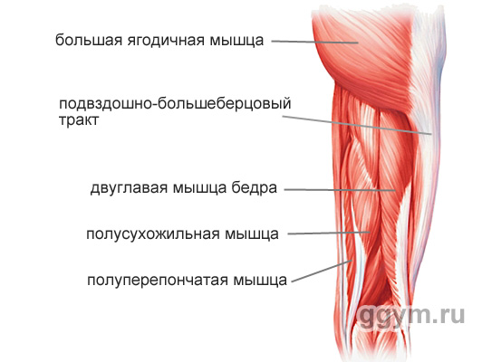
Biceps femoris (m. biceps femoris)
A long, spindle-shaped muscle that runs along the entire back of the thigh. It consists, as the name suggests, of two heads: long and short. The long head is attached at the upper end to the ischial tuberosity of the pelvic bone, and at the lower end to the tibia (tibia). The short upper part is attached to the back surface of the femur, and the lower part is attached to the tibia.

The main functions of the biceps femoris muscle:
Shin flexion (leg bending at the knee)
Hip extension (moving the hip back or straightening the torso from a tilted position)
Maintaining body balance
M. biceps femoris is actively involved in bending the legs, in all movements in which the hip needs to be pulled back, and in extending the body from a tilted position.
Lack of flexibility and strength in the hamstrings is often the cause of back pain, poor posture, and problems with the knee joints.
Semitendinosus muscle (m. semitendinosus)
A long, flat, tapering muscle lying medial (closer to the middle of the body) in relation to the biceps femoris muscle. The upper part of the muscle is attached to the ischial tuberosity of the pelvic bone. The lower one is to the tibia (lower leg).
The main functions of the semitendinosus muscle:
Shin flexion (leg bending at the knee)
M. semitendinosus is actively involved in leg flexion, in all movements in which the hip is required to be pulled back, and in extension of the body from a tilted position.
Semimembranosus muscle (m. semimembranosus)
A long, flat muscle located in the posterior inner part of the thigh. The upper end is attached to the ischial tuberosity of the pelvic bone. The lower end - to various parts of the tibia and fascia of the lower leg muscles.
The main functions of the semimembranosus muscle:
Hip extension (moving it back or straightening the body from a tilted position)
Shin flexion (leg bending at the knee)
M. semimembranosus is actively involved in leg flexion, in all movements in which the hip is required to be pulled back, and in extension of the body from a tilted position.
Muscles of the inner thigh
These muscles are generally called adductors. Their main function is to bring the femur inward.

Thin muscle (m. gracilis)
A long, ribbon-shaped muscle located on top of all other muscles on the inside of the thigh. Its upper part is attached to the pubic bone, and its lower part is attached to the tibia (shin bone).
Main functions of the gracilis muscle:
Shin flexion (bends the leg at the knee)
Rotate the shin inward
M. gracilis is actively involved in all leg movements: running, walking, squatting, maintaining body balance.
Pectineus muscle (m. pectineus)
A flat muscle, attached at the upper end to the pubic bone, and at the lower end to the inner part of the middle of the femur.
Main functions of the pectineus muscle:
Hip adduction (pulls it inward)
Hip flexion (pulls the hip towards the body)
M. pectineus is actively involved in all leg movements: running, walking, squatting, maintaining body balance.
Long adductor muscle (m. adductor longus)
Flat thick muscle. The upper end is attached to the pubic bone, and the lower end to the inner part of the middle of the femur.

The main functions of the adductor longus muscle:
Hip adduction (pulls it inward)
External hip rotation
M. adductor longus is actively involved in all leg movements: running, walking, squats, maintaining body balance.
Short adductor muscle (m. adductor brevis)
A flat muscle that expands downward. Attached at the upper end to the outer surface of the body and pubic bone. The lower (wide end) – to the inner part of the femur.

The main functions of the adductor brevis muscle:
Hip adduction (pulls it inward)
Hip flexion (pulls the hip toward the body, moving it forward)
M. adductor brevis is actively involved in all leg movements: running, walking, squats, maintaining body balance.
Large adductor muscle (m. adductor magnus)
The largest of the adductor muscles, its volume determines the degree to which the space between the thighs is filled. The picture shows the rear view.
Its upper end is attached to the ischial tuberosity of the pelvis and pubic bone. The lower (very widened end) is attached to the inner part of the femur almost along its entire length.

The main functions of the adductor magnus muscle:
Hip adduction (pulls it inward)
Rotates the hip outward
Internal bundles are involved in hip extension (pulling it back and straightening the body from a tilted position)
M. adductor magnus is actively involved in all leg movements: running, walking, squatting, maintaining body balance.
Muscles of the outer thigh
Tensor fascia latae (m. tensor fascia latae)
In general, this is the only muscle, with the exception of the buttock muscles, that is involved in hip abduction.
This is a flat, elongated muscle, tapering downward. The upper end is attached to the anterior iliac spine, and the lower end of this muscle passes into the fascia lata of the thigh - a long tendon that stretches to the lower leg. Being well developed, it gives a pleasant roundness to the lateral surfaces in the pelvic area.
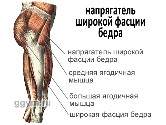
The main functions of the tensor fascia lata are:
Tension of the fascia lata of the thigh (which is necessary for normal leg function when walking and running)
Strengthening the knee joint by stretching the fascia lata
Hip flexion
M. tensor fascia latae is actively involved when walking, running, and performing exercises on one leg.
Well, one last thing worth saying. that the muscles of the thighs and the muscles of the buttocks are interconnected anatomically and according to the functions they perform. A person is characterized by movements in which these muscles work in conjunction: walking, running, squats, bending. As a rule, exercises for developing legs also perfectly develop the buttocks.
The quadriceps muscle performs certain functions in the human body. People can walk, run, and jump largely thanks to muscle tissue.
A question of location
The strongest and most massive muscle in humans is the quadriceps muscle. It is logical to assume that it has 4 components:
- straight;
- medial;
- lateral;
- intermediate.
They all fit the femur. Where the distal third of the thigh is located, thanks to these four heads, the common quadriceps tendon is formed, and it is attached to the kneecap. It then continues down the leg as the patellar ligament. When it encounters the tibia, the ligament attaches to the iliac tuberosity.
The three quadriceps originate on specific areas of the femur:
- 1. External (lateral) - on the outside.
- 2. Internal (medial) - on the inner.
- 3. The intermediate one is located at the front, located between the two previous ones, and is considered the weakest and most vulnerable.
The latter - straight - is distinguished by the fact that its beginning can be traced on the pelvic bone, above the hip joint. It is considered the longest, considering the size of all its heads. Its place is the front surface of the thigh.
Without the quadriceps, a person would look pathetic, or rather, his movements would be very limited. But thanks to the quadriceps muscle structure, the legs at the knee not only move, but also extend. And the rectus femoris muscle, in combination with the iliopsoas, can pull the thigh towards the chest. This is especially appreciated by gymnasts, athletes and those involved in acrobatics and dancing.
But bodybuilders also love to emphasize this muscle; they pump it up to enormous sizes, proudly demonstrating all its grace.
To perform all natural functions, the muscular organs that belong to the thigh do the following work:
- the front ones flex the hip;
- rear - extend;
- medial - adducts the hip.
And since their mass is very large, as well as their length, they have a huge opportunity to develop strength, influencing both the hip and knee joints. The thigh muscles have the fate of performing work of a static and dynamic nature, when a person is both moving and stationary. And they are developed, like the pelvic ones, due to the fact that humans, unlike animals, walk on two limbs.

The quadriceps belongs to the anterior group along with the sartorius muscle structure, which originates from the superior ilium. Its task is to allow the shin to bend, and the thigh to not only bend, but also rotate outward.

Functions performed
Each quadriceps muscle has its own characteristics and purpose.
The straight line has a feathery structure. It begins above the acetabulum, from the inferior ilium.
Between the bone and the beginning of the muscle there is a synovial bursa. Then it continues forward downward from the hip joint, exits the thigh and is located near the sartorius and intermediate muscle structures. Its end consists of a tendon that attaches to the base of the patella.

The largest of all four is the broad lateral one. It begins with both tendon and muscle fibers from the intertrochanteric region and the lower region of the greater trochanter and where the lateral septum is located. Attached to the tendon, which is located in the rectus femoris muscle, and to the tibia. Several of its tendons extend to the lateral patellar tendon.
The origin of the vastus medialis muscle is very large, it covers:
- lower part of the intertrochanteric region;
- medial lip;
- medial femoral septum.
It is attached in the place where the upper edge of the patella is located, and where the anterior part of the medial condyle is designated. As a result, the tendons are directly involved in the process when the vastus medialis patellar ligament is formed to maintain stability.
The vastus intermedius muscle is defined by muscle fibers from the top of the lateralis and femur and where the lateral muscular septum and the lateral labrum area are located.
The quadriceps have large motor units, the content of myosymplasts in them reaches up to 1000.

Myosymplasts are combined into a motor unit, and are part of muscle fibers that have one motor neuron, that is, the ending of nervous tissue that can generate a signal for contraction.


